See details
READ MORE

.jpg)
The biceps femoris muscle is located on the lateral side of the posterior thigh. Together with the semimembranosus and semitendinosus muscles, it makes up the hamstring complex. The biceps femoris is the strongest muscle in the hamstring complex and is responsible for flexion, external rotation, and posterolateral stability of the knee.
The biceps femoris has a long head and a short head. In some cases, the short head of the biceps femoris may be absent. Its long head originates from the ischial tuberosity and the sacrotuberous ligament. The short head of the biceps femoris arises from the lateral lip of the linea aspera, the lateral supracondylar line of the femur, and the lateral intermuscular septum. These two heads of the biceps femoris form the muscle's tendon, which inserts onto the head of the fibula, the crural fascia, and the proximal part of the tibia.
The two heads of the biceps femoris are innervated by different branches of the sciatic nerve. The long head is innervated by the tibial branch of the sciatic nerve, while the short head is innervated by the peroneal branch of the sciatic nerve. This unique innervation of the biceps femoris can lead to a lack of coordination between the two heads and to vulnerability in terms of sports injury.
Injury to the biceps femoris muscle is associated with decreased flexion strength and knee rotational stability. It has been observed that flexion strength decreased by 75% if the biceps femoris tendon is transferred to the fibular collateral ligament. By transferring either part or all of the biceps femoris tendon to the fibular collateral ligament, resistance to unstable anterolateral knee rotators is anticipated, which usually arises due to anterior cruciate ligament insufficiency and injury to the lateral capsule of the knee. On the other hand, it has been shown that the biceps femoris muscle increases its activity in the knee with torn cruciate ligaments. Electromyographic activity of the biceps femoris tendon also increased during quadriceps strengthening exercises and straight-leg raise exercises.
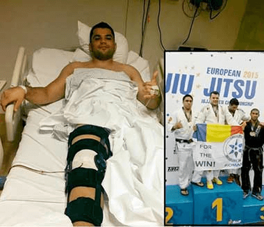
Depending on the severity of the retraction of the biceps femoris tendon, its rupture can be treated either conservatively or surgically. Most patients recover well and are able to return to high-intensity activities, if they receive a timely diagnosis and appropriate treatment.
Rupture of the biceps femoris tendon usually occurs following hyperextension of the knee, concurrent with hip flexion. Soccer is the most common sport associated with biceps femoris tendon injury. In some cases, the injury occurs during hockey, jogging, or water skiing.
Complete rupture of the biceps femoris is rare, and only a few cases have been reported. However, the muscle's tendon is the most frequently affected part of the hamstring complex, since it is the strongest flexor of the leg and an important dynamic stabilizer of the knee.
The biceps femoris muscle becomes active in the late or terminal swing phase of gait. The muscle contracts eccentrically to control the movement of the knee during its extension. The tendon is more vulnerable to injury during eccentric contraction than during concentric contraction. Aging and muscle fatigue make the muscle more susceptible to injury.
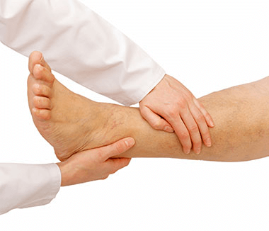
Patients suffering from a rupture of the biceps femoris tendon may complain of sharp pain behind the affected knee during hyperextension. They may feel a pop in the knee while extending it. Most affected individuals had an injury while engaging in activities such as soccer, running, or water skiing. The injury results in the patient's inability to walk. The examiner may palpate the gap next to the ruptured biceps tendon. Sometimes, a subcutaneous hematoma may be palpated. Posterolateral tenderness of the knee is common. Flexion strength of the affected knee may decrease in severe cases.
Imaging examination can help define the extent and severity of the biceps femoris tendon injury. X-rays can be used to check for the presence of a tendon avulsion from the bone, when a bone fragment may be visible. However, in adults, the avulsed bone fragment cannot easily be observed. In contrast, due to incomplete ossification, a displaced bone fragment is easier to observe in adolescents. MRI can be used to assess tendon retraction and the integrity of the surrounding bony structure. Ultrasonography is a relatively inexpensive method and offers the advantage of not exposing the patient to radiation. In addition, this method allows for dynamic assessment of the movement of the biceps femoris tendon relative to the surrounding soft tissues. Doppler ultrasound can be used to assess muscle hypervascularization, which indicates new vascularization or inflammation of the affected tendon.
Treatment of an isolated rupture of the biceps femoris tendon is usually non-surgical. Most athletes can return to competitive sports (professional soccer) within approximately 6 weeks of the injury, if the tendon rupture has a retraction of less than 2 cm. The torn tendon gradually heals, allowing the affected knee to return to its initial strength. Non-surgical treatment includes rest, ice, non-steroidal anti-inflammatory drugs, gentle stretching, and therapeutic exercises for 4-6 weeks. A severe rupture of the biceps femoris tendon may be associated with injury to the semitendinosus and semimembranosus muscles. If tendon retraction is significant (>5 cm), surgical intervention is generally required.
Surgical intervention in this case involves a transverse incision of the gluteal fold, exposing the ruptured tendon, protecting the sciatic nerve, mobilizing the ruptured tendon, and reinserting it onto the ischial tuberosity. Most cases recover well after non-surgical treatment or a timely intervention. There is no indication that specifically addresses when this situation must be treated surgically. Several case studies suggest that a complete rupture of the biceps femoris tendon should be subjected to surgical treatment.
This includes:
Clinicians must be attentive, since not all posterior thigh pain derives from an injury to the biceps femoris tendon. In addition, tendon injury often occurs together with damage to the entire hamstring complex.
The prognosis for biceps femoris tendon injury is good, if diagnosis and treatment are prompt and appropriate. Most athletes return to competitive sports after treatment. Although complete rupture of the proximal biceps femoris tendon is not common, patients may experience weakness, pain, and sciatic nerve irritation if the affected tendon is not treated appropriately.
Education should be made public, since more and more people are taking part in high-intensity exercise. Healthcare providers should offer the best treatments according to evidence-based medicine.
An interdisciplinary approach to biceps femoris tendon rupture is recommended. Rupture of this tendon is relatively common among young athletes and active individuals. Many patients go to the emergency room once the injury has occurred. Therefore, medical staff must know how to diagnose and address this condition. The key to preventing this injury is education about the importance of warming up and stretching before exercise. The prognosis is good for most patients. Non-surgical treatment provides symptom relief and functional recovery. The recommendation for surgical intervention remains uncertain.
Centrokinetic is the place where you will find answers and clear solutions for your movement problems. The clinic dedicated to osteoarticular conditions is divided into the following specialized departments:
Find out the latest news by following the Facebook and YouTube accounts of Centrokinetic clinic.
See details
READ MORESee details
READ MORESee details
READ MORESee here how you can make an appointment and the location of our clinics.








CENTROKINETIC FLOREASCA
Bucharest, 1st District,
Mircea Eliade Street, no. 18,
Eliade Tower, Entrance A, Floor 3
| Navigation | Explore 3D |
CENTROKINETIC Barbu Vacarescu
Bucharest, 2nd District,
Barbu Vacarescu Street, no. 102,
102 The Address, ground floor.
| Navigation | Explore 3D |
CENTROKINETIC MILITARI
Bucuresti, Sector 6,
Bld. Iuliu Maniu, nr. 15G,
Cladirea PBT, etaj 3.
| Navigation |
CENTROKINETIC CLUJ-NAPOCA
Cluj-Napoca,
Str. Intre Lacuri, nr.1
Intrarea este in spatele cladirii.
| Navigation |
SOCRATES CLINIC TIMISOARA
Timisoara, Calea Martirilor 1989,
nr. 1.
Telefon: 0371 785 374.
| Navigation |
Phone: 0319693
E-mail: programari@centrokinetic.ro
Operating hours:
Monday – Friday: 07:00 – 21:00
Saturday: 09:00 – 14:00
Sunday: closed.
MAKE AN APPOINTMENT