See details
READ MORE

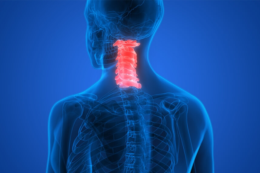
The objective of this study was to measure intradiscal pressure changes in the lower cervical spine during a manual cervical elongation (traction) procedure. Incisions were made anteriorly, and pressure transducers were inserted into each nucleus at the lower cervical discs. Four doctors trained in chiropractic performed the cervical elongation procedure on nine specimens in a prone position, with contacts at C5 or the C6 vertebrae, with the head in different positions.
Changes in intradiscal pressure, traction forces, and manually applied posterior-anterior forces were analyzed using descriptive statistics. Decreases in intradiscal pressure were observed during the manual elongation procedure at all the lower cervical levels, C4-C5, C5-C6, and C6-C7. The average decreases in intradiscal pressure were up to 168.7 KPa (kilopascal). The average traction forces were up to 119.2 N. The posterior-anterior forces applied during manual traction were up to 82.6 N. The decreases in intradiscal pressure were greatest during the flexion and traction movement.
Neck pain and neck-related shoulder and arm pain are a major health problem in Western societies. Symptoms can include pain, tingling, numbness, stiffness, loss of coordination or physical strength, skin discoloration, and temperature differences localized to the neck, shoulder, arm, elbow, wrist, and/or fingers. These complaints cause discomfort and can lead to severe long-term pain and physical disability, creating an economic burden due to work absence and healthcare costs. In 2003, the 12-month prevalence of neck and shoulder pain in the Netherlands was estimated at 31.4% and 30.3%, respectively.
In 2008, approximately 6% of adults in the US reported an outpatient visit for a primary diagnosis for a back or neck condition (13.6 million). Between 1999 and 2008, the average inflation-adjusted annual healthcare expenditures for these patients increased by 95% (from $487 to $950); most of the increase was driven by increased costs for medical specialists, as opposed to primary care physicians. During the study period, average inflation-adjusted annual expenditures for chiropractic care were relatively stable. Physical therapy was the most costly service overall.
Spinal manipulation is used by chiropractors, osteopathic physicians, and physical therapists to treat musculoskeletal conditions. Although spinal manipulation has proven effective in some studies, and researchers have conducted experimental studies with humans and animals, the exact mechanisms behind these techniques are not fully understood.
Cox developed a form of chiropractic manipulation, using a specially designed table, which incorporates traction called manual cervical elongation. Several case studies have reported clinical improvement in patients with neck pain. Manual elongation is hypothesized to create intersegmental motion at a targeted segment during the application of traction through a hand contact that localizes the load using the treatment table. The effects of cervical spine traction can include vertebral separation, reduction of intradiscal pressure, facet joint separation, increase of the intervertebral foramen, and stretching of soft tissues.
It is believed that the intersegmental motion induced by traction opens the intervertebral foramen and decreases intradiscal pressures.
The manual cervical elongation procedure used in the present study is widely used, with 64% of chiropractic doctors treating neck pain with this method.
The objectives of this study were; in unembalmed cadavers with intact head, neck, and trunk: (1) measure intradiscal pressure in the lower cervical spine (C4-C5, C5-C6, C6-C7, and C7-T1) and (2) during the manual cervical elongation procedure performed by the chiropractic doctor, measure the magnitude and reliability of the forces applied.
A specially modified treatment table incorporated a multi-component force plate (3 forces and 3 moments) (model number 2850-06, Bertec, Inc., Columbus, OH) in the thoracic area of the table, on which the torso specimen was mounted (figure 1). The table's head support allowed linear movement to create cervical spine traction, head flexion movement, and locking of the head support at a given flexion angle.
.png)
(Figure 1)
Nine fresh-frozen cadavers, with intact head, neck, and trunk with shoulders, were procured from approved tissue banks and stored in freezers at -20 C. X-rays were taken to rule out severe degeneration, trauma, tumor, or significant osteoporosis.
Figure 2 is a static fluoroscopic video image showing (see arrows) the location of the pressure transducers in the nucleus of the C4-C5, C5-C6, C6-C7, and C7-T1 intervertebral discs. The intervertebral discs were graded from the static fluoroscopic video images by three independent observers using disc height measurements.
.png)
(Figure 2)
The pressure sensors were calibrated using a handheld pressure calibration device (model number HTP1, Druck, Ltd., Leicester, United Kingdom). Pressure calibrations were linear, and a first-order polynomial function was used to describe each calibration. The specimens were thawed at room temperature. Pressure transducers were inserted via anterior approach into the nucleus pulposus at C4-C5, C5-C6, C6-C7, and C7-T1. The transducers were introduced through a 14-gauge cannula into each disc nucleus, under fluoroscopic video guidance (OEC-9800, GE Healthcare Systems, Waukesha, WI).
After the sensors were inserted, the cadaver was placed in a prone position, with the head resting on the mobile head support and the thorax resting on the fixed section of the table, which was mounted on the force plate. The thorax was rigidly fixed to the table section and the underlying force plate by means of Velcro straps. The head and upper cervical spine were positioned on a mobile surface. The thorax was positioned on the middle section of the table, with the cervical spine between the mobile surface and the thoracic section of the table. This allowed manual contact with the vertebrae of the cervical spine and upper thoracic spine for decompression in a neutral position or in a head flexion position.
.png)
The manual elongation treatment protocol used was as follows. The part of the hand between the thumb and index finger was placed on the spinous process and lamina above the segment to be decompressed. (figure 3 (a)). Therefore, a controlled cephalad elongation was applied to the vertebral segment through hand contact and movement of the table support in the longitudinal direction of the spine. Elongation along the length of the spine was applied in three twenty-second elongation stages. Every twenty seconds, there were five loading-unloading cycles (Figure 4). These elongation sessions were applied at the C5 and C6 hand contact locations.
.png)
(Figure 4)
After that, the cervical end of the table was placed at a fixed flexion angle of 15 degrees, and the cervical elongation procedure was repeated while the cervical end of the table moved into flexion and slid longitudinally. The cervical end of the table was allowed to move freely into flexion, while simultaneously sliding along the cranial-caudal axis to create traction. Thus, the doctor simultaneously moved the head piece into flexion and traction, and the cervical spine was subjected to flexion and traction movements simultaneously.
In another procedure, occipital restraints were placed on the cadaver's skull, and the doctor's hand contacted the vertebral arch at the T1 level, with the heel of the hand contacting the spinous process at the T1 level (Figure 3 (b)). Then, generalized traction was applied to the entire cervical spine by moving the table bar in the superior direction. This procedure was repeated with 15 degrees of flexion of the cervical surface of the table.
Figure 4 shows typical graphs of intradiscal pressure at each of the lower cervical discs (C4-C5, C5-C6, C6-C7, and C7-T1) as a function of treatment duration, demonstrating the decrease in intradiscal pressure as the chiropractic doctor applies manual cervical elongation during the five loading/unloading cycles in a given session. Figure 5 shows a typical graph showing the changes in intradiscal pressure as a function of traction force (measured by the force plate under the thoracic support).
.png) | .png) |
  |
(Figure 5)
It is believed that the decrease in intradiscal pressure allows disc prolapse to retract, contributing to improved transport of solutes and nutrients and modifying the chemical environment of the nociceptors in the outer annular layers of the disc. Manually localized lumbar decompression has already been shown to decrease pressure in cadaveric lumbar discs. In addition, its clinical effectiveness for patients with radiculopathy has been demonstrated in a randomized clinical trial. According to doctors (personal communications), the decompressive procedure is commonly used to treat patients with neck pain with radiating symptoms to the arms, where the discs of the lower cervical spine (C4-C5, C5-C6, C6-C7, and C7-T1) are involved.
This study was designed to measure intradiscal pressure changes during a manual elongation procedure in the lower cervical discs (C4-C5, C5-C6, C6-C7, and C7-T1). In this study, longitudinal traction along the length of the spine with contacts at C5 and C6 was performed with the cadaver in a prone position. This position allowed contact with the posterior arch of the specified cervical vertebra (C5 or C6). This is substantially different from standard forms of spinal traction, performed in a seated or standing position, which apply forces to the spine without a localizing contact.
Although the procedures studied can be applied to all patients in the clinical setting, most patients receive one, but not all, of these procedures. Most patients with discogenic pain receive neutral traction or fixed flexion and traction. Few chiropractic doctors use moving flexion and manual elongation combined. Patient tolerance guides the selection of the specific traction procedure. Part of the study was to determine whether there was an additional or varied physiological benefit (decrease in intradiscal pressure) when performing traction alone, or traction with fixed flexion, or combining flexion and traction simultaneously. A two-minute recovery time was allowed between the different traction conditions. Previous biomechanical studies have used recovery times ranging from 15 seconds to 4 minutes. To reduce testing time and tissue degradation, we chose a two-minute recovery time.
Decreases in intradiscal pressure were observed at all levels for DC 1 (doctor of chiropractic) under neutral traction conditions. Intradiscal pressure increased at the C7-T1 level for some of the DCs when contact was at the C6 level. During generalized traction, intradiscal pressure at C7-T1 increased for most doctors. Overall, doctors applied greater forces at the C5 contact compared to C6, and greater forces during the flexion movement compared to neutral and fixed flexion traction. DC4 applied the maximum traction force, while DC3 applied the lowest traction force. DC3 was the clinician in an academic research setting. All four doctors applied posterior-anterior (PA) force together with traction. The level of PA forces was highest for DC4, followed by DC1, then DC2. DC3 had the lowest forces of all the doctors. All doctors applied a greater PA force when contact was at C6. Contact at C6 was more difficult due to the anatomical region, which may explain this finding.
The position and contact force of the hands for each of the doctors were likely different and may have influenced the lordosis of the cervical spine, which, in turn, could have contributed to variations in traction forces. The decrease in intradiscal pressure can be induced not only by the traction forces applied, but also by the traction forces of the intervertebral disc produced by increased lordosis. Thus, while the occurrence of lordosis may have decreased the reliability of the traction force performed by the doctor, the additive effect on intradiscal pressure due to traction and lordosis could improve the reliability of the intradiscal pressure change.
Disc degeneration has an influence on intradiscal pressure changes. In this study, it was found that only two discs had degeneration greater than grade I. Therefore, disc degeneration was not considered as a factor in our observations.
Unembalmed cadavers were used in this study, and it is recognized that active musculature in vivo situations could alter intradiscal pressure. It is standard practice in numerous biomechanical studies published in the literature to use human spine specimens to assess the mechanical response of intervertebral discs, to understand how the human spine can respond to physiological loads (forces and moments) experienced during daily activities.
In this cadaveric study, decreases in intradiscal pressure were observed in the lower cervical spine during a chiropractic manual cervical elongation procedure in the prone position. Based on the maximum number of specimens the doctors performed, the flexion and traction movement appears to reduce intradiscal pressure the most, followed by neutral traction, fixed flexion and traction, and generalized traction. Although the chiropractic doctors in this study demonstrated good intra-clinician reliability, the magnitude of the traction forces varied. Higher-powered studies should be undertaken to determine whether these decreases in intradiscal pressure are significant depending on the doctor, the contact location, and the different traction procedures. The clinical significance of these differences is also not known.
Centrokinetic is the place where you will find answers and clear solutions for your movement problems. The clinic dedicated to osteoarticular conditions is divided into the following specialized departments:
Find out the latest news by following the Facebook and YouTube accounts of Centrokinetic clinic.
See details
READ MORESee details
READ MORESee details
READ MORESee here how you can make an appointment and the location of our clinics.








CENTROKINETIC FLOREASCA
Bucharest, 1st District,
Mircea Eliade Street, no. 18,
Eliade Tower, Entrance A, Floor 3
| Navigation | Explore 3D |
CENTROKINETIC Barbu Vacarescu
Bucharest, 2nd District,
Barbu Vacarescu Street, no. 102,
102 The Address, ground floor.
| Navigation | Explore 3D |
CENTROKINETIC MILITARI
Bucuresti, Sector 6,
Bld. Iuliu Maniu, nr. 15G,
Cladirea PBT, etaj 3.
| Navigation |
CENTROKINETIC CLUJ-NAPOCA
Cluj-Napoca,
Str. Intre Lacuri, nr.1
Intrarea este in spatele cladirii.
| Navigation |
SOCRATES CLINIC TIMISOARA
Timisoara, Calea Martirilor 1989,
nr. 1.
Telefon: 0371 785 374.
| Navigation |
Phone: 0319693
E-mail: programari@centrokinetic.ro
Operating hours:
Monday – Friday: 07:00 – 21:00
Saturday: 09:00 – 14:00
Sunday: closed.
MAKE AN APPOINTMENT