See details
READ MORE


Bone marrow edema (BME) is a descriptive term for a common finding on magnetic resonance imaging (MRI). It can occur in almost all bones, but is most often observed in the lower extremities. BME can be symptomatic or asymptomatic. Therefore, painful BME must be differentiated from incidental BME. This article covers the diagnosis and treatment of painful BME. Painful BME can occur spontaneously (primary BME: BME Syndrome (BMES)) or secondary to various diseases covering almost every medical specialty (secondary BME). Consequently, differentiating between primary and secondary BME, as well as identifying the underlying condition in secondary BME, is of the utmost importance for initiating appropriate treatment. However, evidence-based interdisciplinary guidelines for the diagnosis and management of BME are lacking.
This is problematic, since patients suffering from painful BME are seen by various different medical professions, regardless of whether their clinical background covers the underlying pathology. In addition, different medical specialties often follow their own diagnostic path. In the absence of interdisciplinary management algorithms, this frequently leads to repeated diagnostic measures (resulting in additional healthcare costs), an unsatisfactory patient journey, incorrectly treated or delayed diagnosis, and insufficient treatment with potentially harmful consequences. The lack of an evidence-based interdisciplinary diagnostic algorithm led us to initiate a study and a concrete diagnostic and treatment plan, with the aim of developing diagnostic algorithms based on available evidence and expert opinion.
This algorithm was presented at various national meetings, critically discussed, and adapted. The results were redefined, summarized, and ultimately used as a standard operating procedure.
In this article, we present our algorithm for the evaluation and management of BME, together with a comprehensive overview of the different pathologies/diseases that could underlie BME. This is not a guideline or recommendation from a national medical society, but rather a single-center perspective, with recommendations derived from a review of the literature and practical experience, which can be critically discussed and adapted to other facilities. However, to our knowledge, it is the first published perspective on the interdisciplinary management of BME in Romania. This may help improve the quality of care for these patients until higher-quality evidence-based guidelines are published.
Although the modified signal pattern of BME observed on MRI images is likely related to a modification of normal bone marrow by a material richer in water or increased tissue vascularization, the actual histopathological mechanism of BME remains unknown. To address this subject, we conducted an in-depth literature review to identify studies that investigated the underlying molecular, immunological, and histopathological findings in BME from various causes. These studies are summarized and grouped by their aspect. In summary, studies investigating inflammatory causes of BME, such as spondyloarthropathy, ankylosing spondylitis, or rheumatoid arthritis, show increased vascularization and cellularity, consisting mainly of immune system cells (T cells, B cells, macrophages). In contrast, studies on BME in advanced osteoarthritis have shown reduced perfusion, thickening of the subchondral plate, and increased bone resorption. Osteonecrosis somewhat reflects the pathological changes observed in osteoarthritis. However, these histological and molecular changes are not related to the subchondral bone. Studies on BMES are generally inconclusive and, based on the criteria defined in this document, do not always meet the level of diagnostic accuracy required for diagnosing BMES. Overall, data for any BME entity are scarce and are mainly limited to situations where BME is already present. Further analyses of the underlying molecular mechanisms are needed to provide new pathophysiological insights that could lead to new disease-specific therapeutic targets.
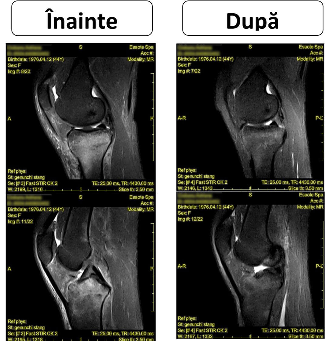
MRI is an indispensable tool for detecting or excluding various differential diagnoses for BME. The sequence protocol for BME should include fat-saturated proton density sequences, such as PDW FS, in three spatial orientations, as well as an unenhanced T1w sequence and a fat-saturated T1w sequence after gadolinium administration. BME can be detected after gadolinium administration; BME shows an early and strong enhancement, while necrotic tissues typically do not show contrast enhancement. A more sensitive approach for differentiating BME from osteonecrosis is high-temporal-resolution perfusion imaging, with quantitative determination of plasma flow and mean transit time.
Traumatic BME resulting directly or indirectly from trauma includes traumatic fracture BME (or micro-fracture), post-surgical BME, and complex regional pain syndrome. Depending on the force, load transmission, and bone quality, trauma initially causes an injury/fracture of the trabecular bone microarchitecture (traumatic BME), which can progress to cortical fractures (typical fracture) and fracture dislocation. Trabecular disruption causes an increase in the fluid level detected by MRI. The affected region (including both adjacent joints) must be immobilized, and the patient is advised not to bear any weight on the affected extremity.
Septic BME. Warning signs for septic BME (osteomyelitis/osteitis, septic arthritis) are age >80 years, implants, a recent injection or surgical intervention, intravenous drug abuse, skin lesions, and immune defects, including diabetes.
Primary inflammatory/rheumatic BME in rheumatology. BME is of great importance for the diagnosis of musculoskeletal inflammation, including arthritis, spondylitis, and enthesitis. Subchondral BME lesions in arthritis result from osteitis characterized by infiltrates of lymphocytes, plasma cells, and macrophages that are linked to osteoclasts replacing the fatty bone marrow BME; they can already be observed a few weeks after symptom onset and attenuate after effective anti-inflammatory treatment. Subchondral BME is a strong predictor of subsequent joint destruction.
In axial spondyloarthritis (axSpA), BME in the spine and sacroiliac joints is considered an early sign of axial inflammation and is associated with histological inflammation, clinical symptoms, and radiographic progression.
Mechanical/degenerative-type BME includes osteoarthritis, insertional tendinopathies, osteochondral lesions, and bone stress injuries. Osteoarthritis (OA) is no longer considered a cartilage degeneration pathology, but rather a combination of pathologies that includes the synovium and subchondral bone. MRI and histopathological examinations have shown that BME in osteoarthritis is a combination of bone marrow fibrosis and necrosis rather than edema, and should therefore be called bone marrow lesion (BML) rather than BME. BML has been associated with pain and is predictive of joint replacement intervention. BML can be considered a possible target for future OA treatment strategies. Several RCTs have shown that bisphosphonate-based therapy resulted in a considerable reduction in the size and pain experienced by the patient due to BML and OA. However, validated data showing a positive effect on the progression of OA are lacking.
Stress-induced bone injuries occur after increased-intensity forces are applied repetitively, or after atypical forces due to joint instability. Microfractures present as BME on an MRI examination, as well as their accumulation, show us that stress-induced fractures can result. Although stress-induced bone injuries can occur in any bone, injuries occurring in the lower limb (40%) and foot (35%) are the most common. The key to successful treatment of this pathology lies in early diagnosis and identifying the nature of the injury (stress injury, stress reaction, stress fracture, or instability).
Neoplastic. Although very rare, neoplastic causes must be considered when evaluating BME. Solid tumors can induce reactive bone marrow edema. Primary bone cancer is rare. The most common are osteosarcomas, chondrosarcomas, and Ewing sarcoma, which usually occurs in children. The only benign, painful bone lesion that can mimic BME is osteoid osteoma, which is common in children or adolescents. Hematological neoplasms can lead to suspicious-appearing lesions on MRI examination, including BME. Acute leukemias or chronic myeloproliferative neoplasms can be diagnosed through blood cell examination. Pathological fractures and osteolysis are typical manifestations of multiple myeloma, which affects the bone marrow throughout the body.
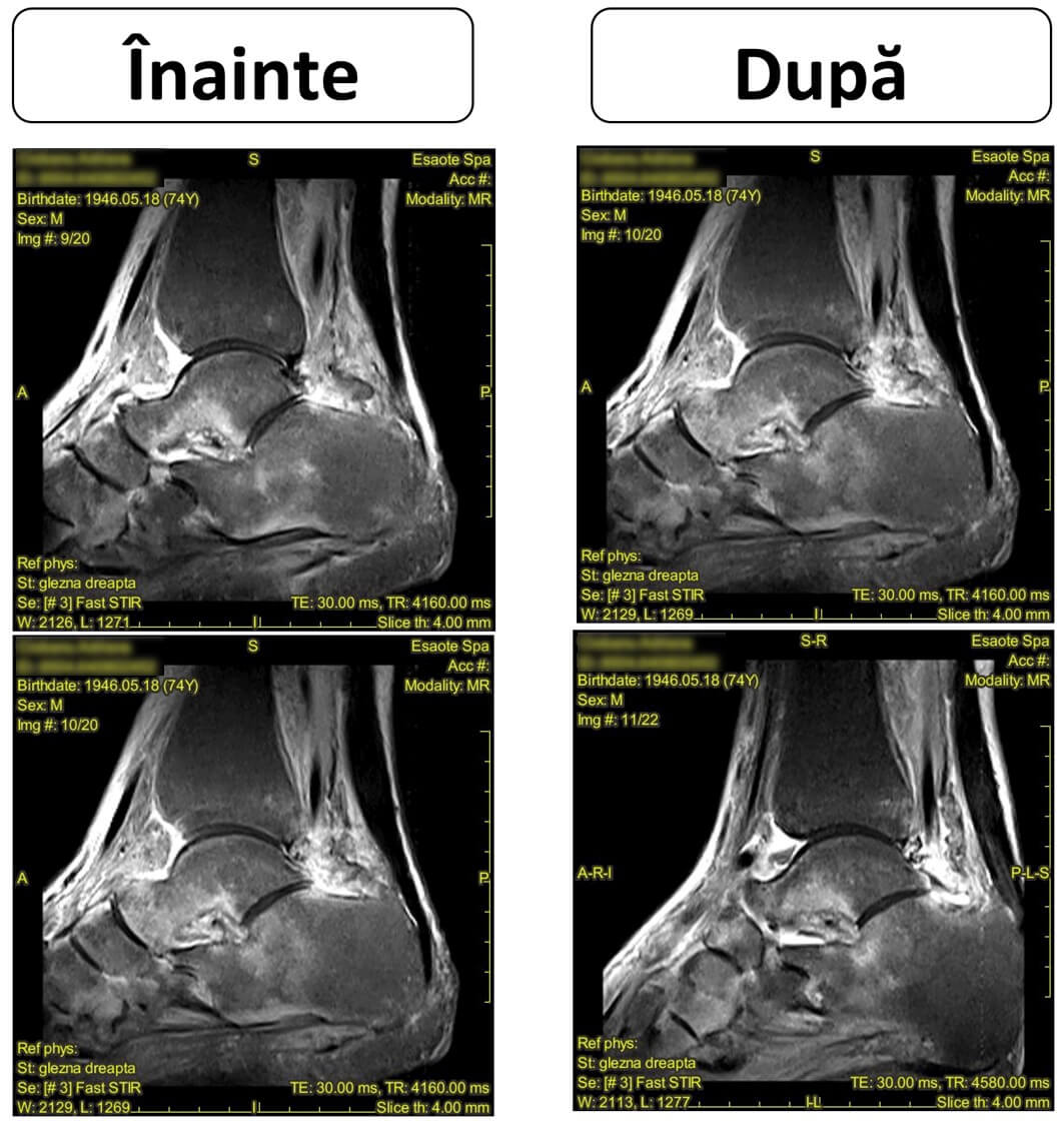
Ischemic BME comprises avascular osteonecrosis and Charcot neuro-osteoarthropathy. Diagnosing avascular necrosis (AVN) can be challenging and results from CT or MRI imaging. AVNs can be classified according to the ARCO criteria. Although BME is often considered an early sign of AVN onset, recent studies have highlighted the fact that BME only appears in advanced stages of the disease, specifically in ARCO stages III and IV, as a symptom of biomechanical deterioration of the trabecula. AVN can occur in the absence of trauma or an impending fracture with vascular rupture. The exact pathophysiology of atraumatic AVN is unclear, but numerous risk factors have been identified, including glucocorticoid use, alcohol consumption, trauma, chemotherapy, kidney transplant, or blood clotting abnormalities, which can also include sickle cell disease.
Metabolic BME. If this is not the case for painful BME, it may be found during the course of a prior diagnostic workup, or it may indicate a refractory, recurrent, and/or migratory factor, a metabolic one (primary/secondary osteoporosis), which should be highlighted. Vitamin D deficiency is common, especially among the elderly population. In contrast, secondary hyperparathyroidism with severe osteomalacia and typical pseudofractures (Looser's zones) are rather rare in developed countries. Current guidelines recommend moderate and minimal avoidance of Vitamin D deficiency in patients who are in the risk category for developing microfractures. A similar partial mineralization condition is adult hypophosphatasia (aHPP). Clinical signs occurring in adults include musculoskeletal pain and fragility fractures, especially metatarsal, femoral, and stress fractures.
The above-mentioned secondary causes of BME can also be considered possible causes for the occurrence of secondary osteoporosis. Osteoporosis is defined as an increase in the degree of risk for the occurrence of microfractures, which can further progress into secondary BME. Cross-sectional studies suggest that the overall prevalence of osteoporosis is a substantial risk for the occurrence of BME. Terms such as transient, migratory, or regional osteoporosis should be avoided, given that they have been used inconsistently to determine the causes of metabolic disease and BME. It should be noted that the diagnostic sensitivity of DXA is low, with more than 50% of patients suffering a typical fragility fracture showing normal or osteopenic DXA results.
Diamagnetic therapy is a non-invasive procedure that involves the use of both high-intensity and low-intensity magnetic field pulses.
The physical principle of the diamagnetic procedure is based on recognizing the effects of the magnetic field on biological tissues, during the use of a magnetic field starting from a low frequency, on several substances such as water or the proteins that make up the extracellular or intracellular membranes.
This targets:
All these programs have positive effects on the recovery of musculoskeletal pathologies.
This device generates electromagnetic pulses marked by a low frequency of 7Hz and a high one of 2.2 Tesla. This device was designed to be used for the medical purpose of diamagnetic therapy and to exploit the physical principles of magnetic waves or diamagnetic molecular acceleration; furthermore, the technology is built on an energy transfer system in both capacitive and resistive modes. The additional energy capacity reaches up to 90 Joules, while the device's safety profile guarantees that it belongs to the non-radiation class.
Operating modes:
This involves the use of frequencies below 300 KHz, the interaction between exogenous impulses and the electrical activity of the nerve endings, and can be improved by selecting frequencies according to the type of pain and the strength of the magnetic field. The electrical impulses sent by the magnetic field interfere with the activity of the nociceptors of the nerve fibers involved in transmitting the sensation of pain; thus, at a frequency of 215 KHz, the electrical stimuli charge the nociceptors, thereby inhibiting the propagation of pain.
Endogenous biostimulation of tissues depends on the characteristics of the electric field induced at the cellular level.
The diamagnetic pump detects the frequencies emitted by the tissue, thus rearranging the electrical potential of the cell membrane altered by the pathological state. The induced electric field is endogenous because it is generated inside the tissue, and isotropic because it has a homogeneous distribution in the treatment area.
The transduction of the electrical signal into biological activity triggers:
In the human body, molecules such as water and many of the proteins and ions, once they enter the high-intensity magnetic field, generate a magnetic force in the opposite direction. The boomerang effect is specific to the CTU 20, thus rebalancing the activities of the cells' metabolic system, moving and shifting cells and solutions within the extracellular matrix and the intracellular components.
The extracellular volume is effective in post-traumatic processes, inflammations, and lymphatic and vascular edemas, while the intracellular volume acts on the cell's cytoplasmic component, thus restoring the cells' enzymatic functions (ATP production, protein synthesis).
The parameters differ according to the proportions of fluid movement and are set according to:
Diathermy allows physical energy to be delivered in both capacitive and resistive modes, generating a thermal effect, vasodilation, and hyperemia, all of which together improve the flow of fluids and solutions necessary for the cells' metabolic activity and ensure a hydrostatic balance and colloid-osmotic pressure.
The effects of diathermy, combined with those of diamagnetotherapy, offer the advantage of increasing fluid drainage.
The CTU Mega20 diamagnetic pump has the ability to explore the electrical impedance of each tissue, assessing the changes for each selected program.
Centrokinetic is the place where you will find answers and clear solutions for your movement problems. The clinic dedicated to osteoarticular conditions is divided into the following specialized departments:
Find out the latest news by following the Facebook and YouTube accounts of Centrokinetic clinic
See details
READ MORESee details
READ MORESee details
READ MORESee here how you can make an appointment and the location of our clinics.








CENTROKINETIC FLOREASCA
Bucharest, 1st District,
Mircea Eliade Street, no. 18,
Eliade Tower, Entrance A, Floor 3
| Navigation | Explore 3D |
CENTROKINETIC Barbu Vacarescu
Bucharest, 2nd District,
Barbu Vacarescu Street, no. 102,
102 The Address, ground floor.
| Navigation | Explore 3D |
CENTROKINETIC MILITARI
Bucuresti, Sector 6,
Bld. Iuliu Maniu, nr. 15G,
Cladirea PBT, etaj 3.
| Navigation |
CENTROKINETIC CLUJ-NAPOCA
Cluj-Napoca,
Str. Intre Lacuri, nr.1
Intrarea este in spatele cladirii.
| Navigation |
SOCRATES CLINIC TIMISOARA
Timisoara, Calea Martirilor 1989,
nr. 1.
Telefon: 0371 785 374.
| Navigation |
Phone: 0319693
E-mail: programari@centrokinetic.ro
Operating hours:
Monday – Friday: 07:00 – 21:00
Saturday: 09:00 – 14:00
Sunday: closed.
MAKE AN APPOINTMENT