See details
READ MORE

Centrokinetic is the leading medical clinic dedicated to bone and joint conditions in Bucharest, having the following medical specializations: orthopedics, neurology, medical imaging, medical rehabilitation, rheumatology, nutrition and psychology, dedicated to both adults and children.
Designed, created and led by one of the most renowned orthopedic doctors in Romania, Dr. Andrei Ioan Bogdan, the clinic is based on a unique concept of obtaining an accurate diagnosis as quickly as possible through high-performance investigative methods (MRI, musculoskeletal ultrasound, EEG, EMG, bone densitometry), and initiating a complete treatment until the patient is cured.
The Centrokinetic team is made up of doctors highly specialized in bone and joint conditions. What sets us apart from other systems is the method of medical collaboration and the protocols we use, so that all the doctors on the team use the same treatment algorithms, implemented from the most prestigious clinics in the world.
Our team, throughDr. Pantu Cosmin, radiologist specialist and senior lecturer at the Carol Davila University of Medicine and Pharmacy, presents an article about knee anatomy.
The patellofemoral joint is frequently referred to as the extensor mechanism. Although it is true that the concentric action of this motor unit is knee extension, functionally, the quadriceps acts eccentrically during walking, running or jumping. Although it receives less attention during treatment, the extensor mechanism can be involved in cases of knee dislocation, such as a fracture of the lateral femoral condyle through lateral retinaculum avulsion, which occurs in a posterolateral dislocation, or in injuries to the m. vastus medialis obliquus and vastus medialis, which can occur concomitantly with other capsular ligament injuries. In addition, knowledge of the injury response of the retro-quadricipital or infrapatellar Hoffa fat pad helps prepare the surgeon to successfully deal with the secondary sequelae of a knee dislocation.
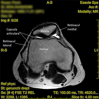
Fig. 1. JOINT CAPSULE
Transverse section, MRI FSE PD.
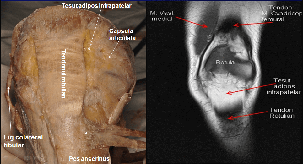
Fig. 2. PATELLAR TENDON - anterior view
Dissection image showing the appearance of the patellar tendon from an anterior view. We note the two insertions, patellar and tibial. Distally it is separated from the tibial tuberosity by the infrapatellar bursa. The patellar tendon comprises a superficial layer, which is contiguous with the retinacular layer, and a deeper layer, which is considered part of the extensor mechanism of the knee. These layers are largely adherent, similar to the insertion of the subscapularis tendon on the anterior capsule of the shoulder joint.
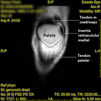
Fig. 3. PATELLAR TENDON - normal MRI appearance of the proximal insertion
Coronal MRI section showing the proximal insertion of the patellar tendon
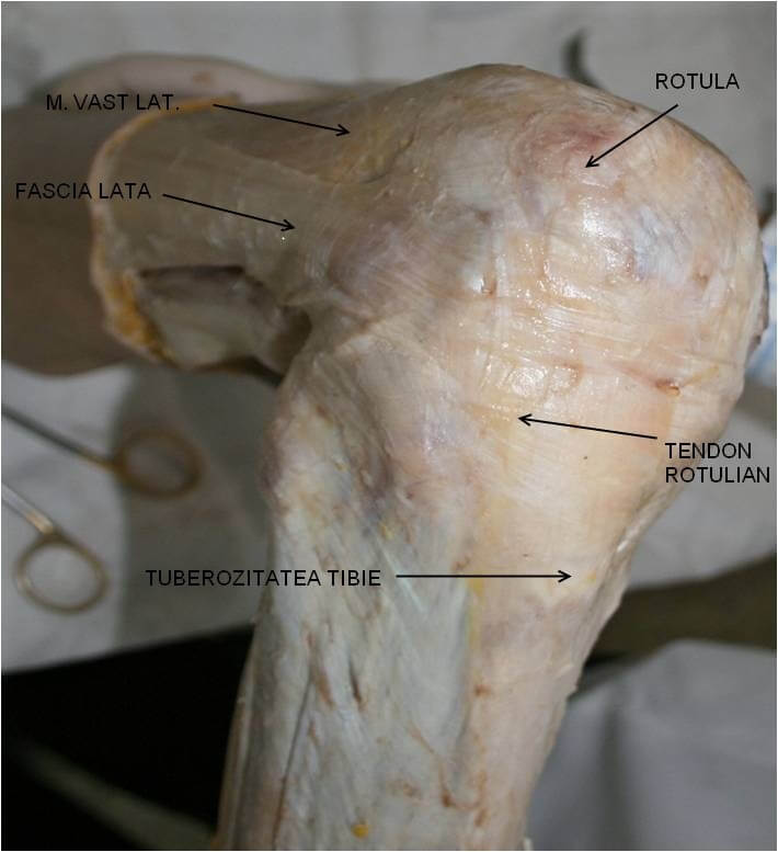
Fig. 4. PATELLAR TENDON – dissection - lateral view
From both the anatomical and imaging pictures, we can observe the patellar ligament as a strong fibrous formation (5-6 cm long and 2-3 cm wide), situated anterior to the joint, with a triangular shape that inserts through its base onto the tip of the patella, and through its tip onto the tibial tuberosity.
It has two surfaces: anterior, corresponding to the femoral fascia, and posterior, corresponding to the infrapatellar fat pad and the deep infrapatellar bursa.
During walking, in which internal rotation occurs, the patellar tendon becomes more vertical, bringing it into the same direction as the quadriceps muscle.
The quadriceps muscle group forms the primary motor unit of the extensor apparatus. The deepest one, the m. articularis genu, is the only fibrillar bundle that does not insert directly into the patella but instead terminates superiorly on a synovial fold, lifting and protecting the patella from possible impingement. The vastus intermedius has a broad origin on the anterior surface of the femoral shaft and descends parallel to the femoral axis. Its distal tendon, separate from the rectus femoris tendon, situated more superficially and separated from it by a discrete bursa, comprises the deep layer of the aponeurosis in this area and inserts directly onto the superior pole of the patella. The m. rectus femoris has a superficial course parallel to the m. vastus intermedius, but terminates in the retinacular layer. The rectus femoris, together with the vastus medialis obliquus, vastus medialis, vastus lateralis and oblique vastus lateralis, terminates in an aponeurosis that merges with the joint capsule. This layer terminates and is adherent to the superficial surface of the patella and continues distally, investing and encompassing the superficial portion of the patellar tendon, eventually becoming contiguous with the tibial periosteum.
And during extension, the patellar tendon is tensioned by the quadriceps muscle, but is relaxed in an upright position. In symmetric standing, the passive mechanisms described above maintain posture with minimal muscular effort, active contraction of the patellar tendon occurring only in asymmetric postures (bending forward, lifting weights).
https://centrokinetic.ro/i.php?p=fig.%2013.jpghttps://centrokinetic.ro/i.php?p=fig.%2013.jpg
Fig. 5. PATELLAR TENDON - MRI imaging appearance - lateral view
MRI FSE PD section. Lateral view of the patellar tendon.
We note the two insertions, patellar and tibial.
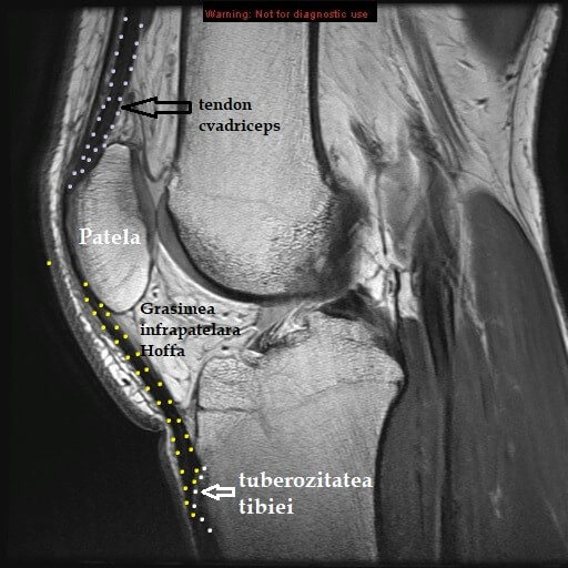
Fig. 6. The patellar tendon
The patellar tendon presents as a strong fibrous formation, 5-6 cm long and 2-3 cm wide, situated in front of the joint. It has the shape of a triangle and inserts through its base onto the tip of the patella, and through its tip onto the lower part of the tibial tuberosity. Since it is considered the terminal tendon of the quadriceps muscle, the patella would in this case be a sesamoid bone.
.png)
Fig 7. MEDIAL AND LATERAL RETINACULA
The patellar wings represent two fibrous bands stretched in a horizontal plane and covered by the quadriceps expansion, extending between the edges of the patella and the femoral condyles.
The retinacula can be compared to rails guiding the patella, with the wings acting as reins.
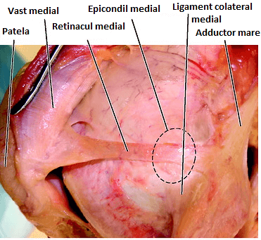
Fig. 9. MEDIAL RETINACULUM
The medial patellar retinaculum, or medial patellofemoral ligament, is part of the medial stabilizing apparatus of the knee. It originates in an area located between the medial epicondyle of the femur and the adductor tubercle, superior to the origin of the superficial component of the medial collateral ligament. The patellar attachment occupies 2/3 of the proximal patellar edge, though sometimes it can extend over the entire patella. The proximal insertion of the retinaculum extends onto the quadriceps tendon, while distally, the ligament passes deep to the oblique fibers of the m. vastus medialis, which also inserts onto the medial edge of the patella. The medial patellofemoral ligament also inserts into the aponeurosis of the vastus intermedius and medialis muscles. This complex insertion creates an anatomically important aponeurosis for the stability of the patellofemoral joint, as it facilitates a dynamic system that guides and pushes the patella into the trochlear groove during active flexion.
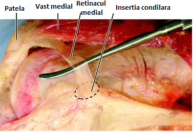
Fig. 10. MEDIAL RETINACULUM - lateral view
We can observe the relationship of the retinaculum with the tendon of the vastus medialis muscle, which has a separate fibrillar expansion called the vastus medialis obliquus. This represents the most important stabilizer of the retinaculum.
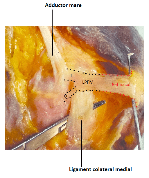
Fig. 11. MEDIAL RETINACULUM - femoral insertion
Anatomically, the transverse band of the medial patellofemoral ligament (MPFL) is also a component of the medial retinaculum, but for the purpose of MRI interpretation, the general convention is to describe abnormalities of the transverse band as MPFL injuries, while more distal injuries involving multiple layers are generally referred to as abnormalities of the medial retinaculum. We note the insertion of the transverse segment of the MPFL together with the fibers of the adductor magnus in the area of the adductor tubercle. There is also a description of an oblique fibrillar bundle that crosses the superficial component of the medial collateral ligament. We can thus observe that the femoral insertion does not occur at a single point but rather over an area that can be extremely variable in location and extent.
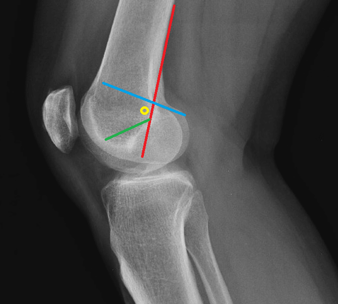
Fig. 12. RADIOLOGICAL LANDMARK OF THE FEMORAL INSERTION OF THE MEDIAL PATELLOFEMORAL LIGAMENT
On a conventional radiograph in lateral view, the condylar insertion of the retinaculum, or Schottle's point, can be identified. This is located at the intersection of two imaginary lines, one following the posterior contour of the femoral diaphyseal cortex (red line), the second being represented by the line of the posterior margin of the medial condyle (blue line) or by Blumensaat's line (green line). The insertion (yellow circle) is located a few millimeters anterior to the intersection of these landmarks.
The medial retinaculum is best visualized on axial or oblique sections, being described as a hypointense band extending posteromedially from the level of the medial edge of the patella toward the outer surface of the medial condyle in the vicinity of the adductor tubercle. At its patellar insertion, the retinaculum is largely thickened due to the fibrillar contribution from the vastus medialis muscle and the sartorius muscle. Toward its femoral insertion, the cross-sectional appearance of the ligament is bilaminar, with two layers evident, one superficial and one deep. This portion of the ligament is, as mentioned, called the medial patellofemoral ligament, part of the medial retinaculum.
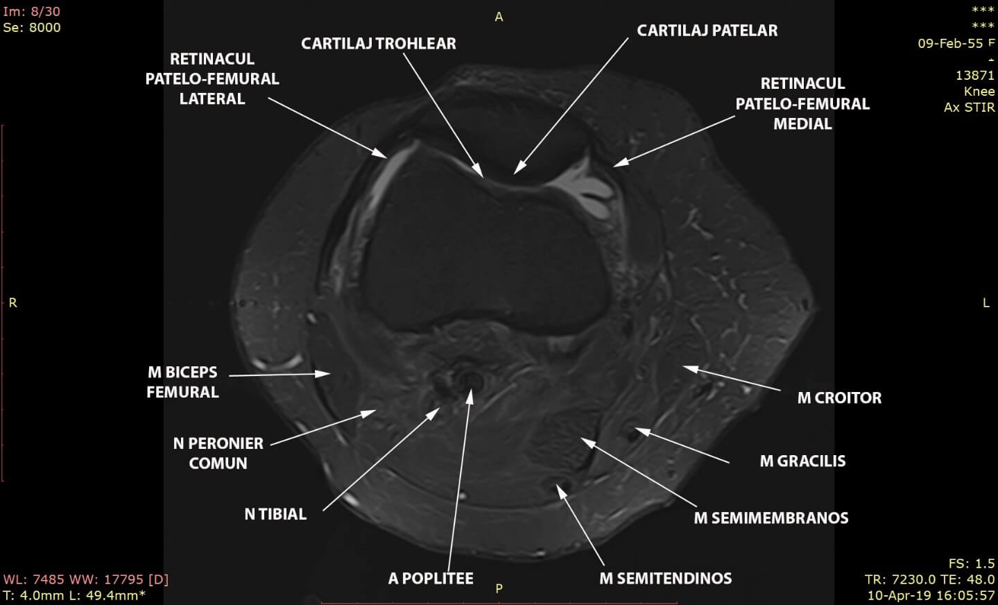
Fig. 13. Transverse MRI STIR section (with fat signal suppression) showing the insertions and course of the two patellar retinacula
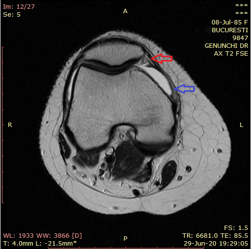
Fig. 14. T2 FS MRI image showing the different appearance of the two segments of the retinaculum
We can observe that the anterior portion, corresponding to the patellar insertion (the retinaculum proper, marked by the red arrow), has a well-organized, dense fibrillar appearance, compared to the posterior, femoral portion (blue arrow), where we identify the two bands, superficial and deep, that make up the medial patellofemoral ligament.
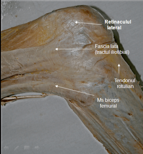
Fig. 15. LATERAL RETINACULUM - dissection views
The joint capsule is perforated anteriorly by the patella, as we can observe, at whose edges it also inserts. The quadriceps expansion represents the fibrous sheet that detaches from the tendons of the quadriceps muscle, then passes like a curtain in front of the knee joint and attaches to the tibia. This presents a series of vertical fibers, on the edges of the patella, which actually constitute the medial and lateral retinacula.
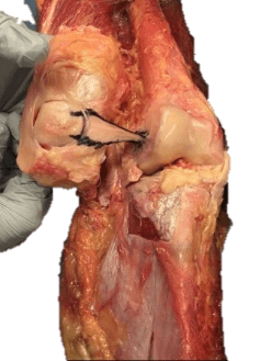
Fig. 16. LATERAL RETINACULUM - intra-articular view
The lateral patellofemoral ligament shown on a cadaver. The patella is reflected laterally, thus revealing the intra-articular appearance of the lateral retinaculum.
In order to show the lateral retinaculum, we used a lateral approach at the level of the iliotibial tract. This was detached at its insertion on Gerdy's tubercle and reflected proximally. This reveals a tendinous thickening extending between the lateral epicondyle of the femur and the lateral edge of the patella. In the next step we used a contralateral parapatellar approach, dissecting the patellar tendon and the quadriceps muscle in order to obtain both an intra- and extra-articular view. On palpation, the ligament was identified as a discrete thickening of the lateral portion of the capsule, with well-defined superior and inferior margins. Through careful dissection of the synovium, we were able to isolate the lateral retinaculum.
.png)
Fig. 17. LATERAL RETINACULUM - lateral view
We observe the oblique course of the lateral patellar retinaculum between its two insertions.
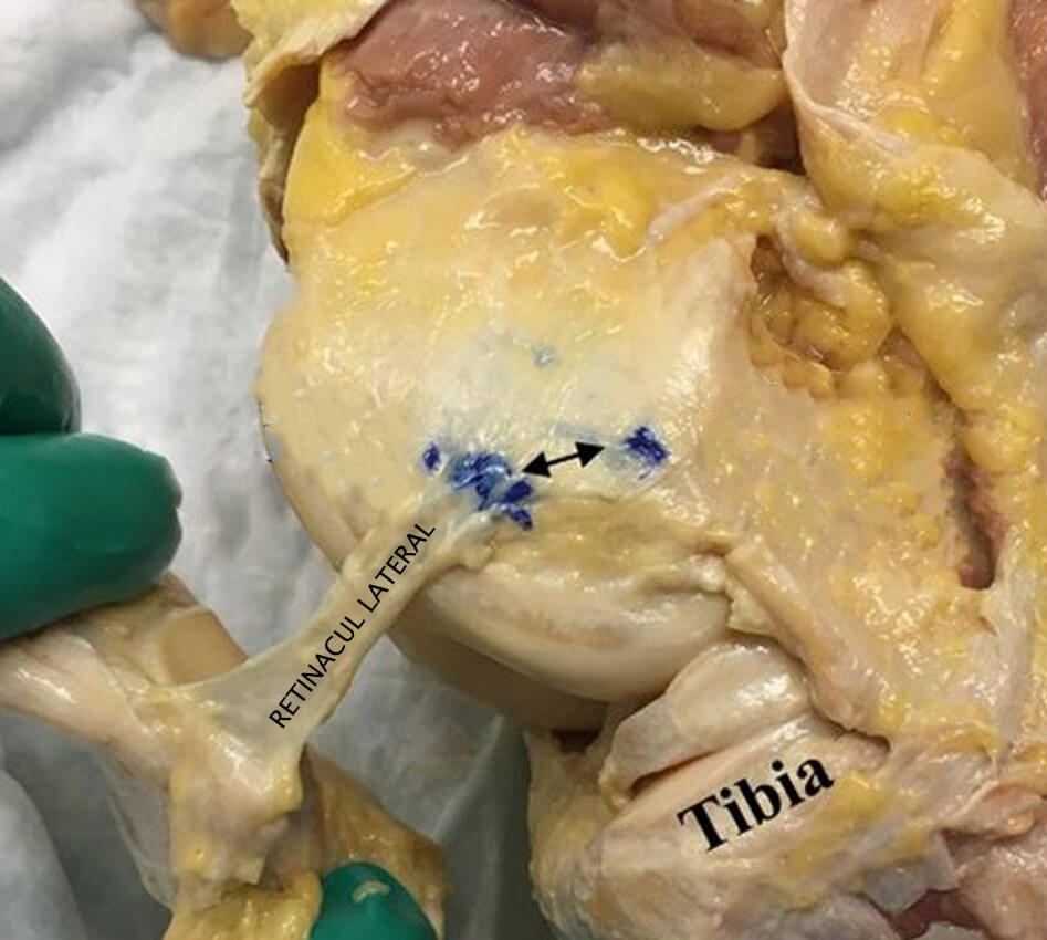
Fig. 18. LATERAL RETINACULUM - femoral insertion
We note the distance between the insertion of the retinaculum and that of the fibular collateral ligament.
.png)
Fig. 19. PERIARTICULAR MUSCLES
We can conclude that any adopted extension position represents a balance between the forces that extend the joint (quadriceps muscle and patellar tendon) and the opposing passive mechanisms (cruciate ligaments, collateral ligaments, the posterior region of the capsule, the posterior oblique ligament, the fascia and the skin).
It should be emphasized, however, that the described capsulo-ligamentous elements have a limited passive mechanical resistance, lower than what is required by knee mechanics, and the factors that truly stabilize the knee are the periarticular muscles. Muscles that come into action through the information received from the proprioceptive receptors that abundantly populate the knee ligaments. This explains the limited effectiveness, both in absolute terms and over time, of replacing torn natural ligaments through ligamentoplasty.
Patellar retinacula pathology is closely correlated with patellar instability and subluxation.
The first episode of patellar dislocation usually occurs after knee sprains, at which point the medial ligamentous stabilizers tear, and the patella strikes the lateral femoral condyle. The typical injury pattern usually involves a tear of the medial patellofemoral ligament, as well as the presence of bone contusions of the patella and the lateral femoral condyle. In addition, complex injuries to the bone segments, cartilage or other knee ligaments may occur. This first subluxation favors future patellar dislocations, especially if additional risk factors are present.
Recurrent patellar dislocations usually occur in individuals with anatomical variants of the patellar stabilizers, such as trochlear dysplasia, patella alta, and lateralization of the tibial tuberosity. Magnetic resonance imaging (MRI) is reliable in identifying risk factors for chronic patellar instability and in evaluating knee joint injuries associated with patellar dislocation.
MRI imaging can thus provide important information for individual treatment. Patients with primary patellar dislocation, without serious internal knee injuries, who do not have major risk factors, can be treated conservatively.
Patients with severe ligamentous tears or large osteochondral lesions require prompt surgical intervention. In addition, surgical correction of anatomical variants will help reduce the potential for chronic instability.
The most common procedures, besides retinaculum reconstruction, include trochleoplasty, tibial tuberosity medialization, and medial capsular plication.
For a comprehensive evaluation of patellar dislocation, a radiologist must be able to identify typical injury patterns, know the standard methods for evaluating risk factors for patellar instability, and be familiar with surgical options.
Most patients with patellar dislocation are young, active people, with women in the second decade of life having a higher risk. Nearly half of all patients with a first-time dislocation will experience subsequent dislocations after initial conservative treatment. During the recovery period, most patients have reduced mobility, and two-thirds of them report limitations in difficult activities. Chronic instability of the patellofemoral joint and recurrent dislocation can lead to progressive cartilage deterioration and later to the onset of severe arthritis, if not treated appropriately.
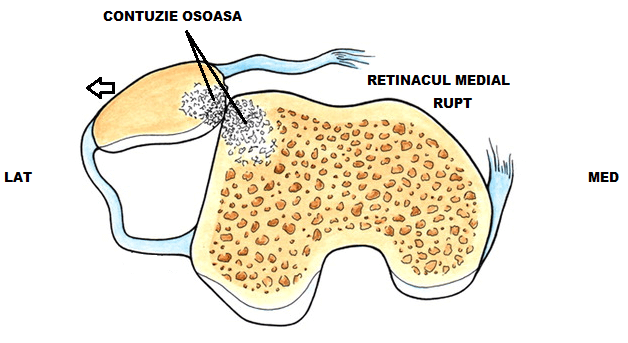
Fig. 20. MECHANISM OF TRAUMATIC PATELLAR SUBLUXATION - diagram
Trochlear dysplasia has been identified as one of the main factors contributing to chronic patellofemoral instability. In individuals with trochlear dysplasia, the surface of the trochlear joint is flattened proximally, and the concavity is less pronounced distally. This combination results in considerable loss of patellar contact and lateral dislocation of the patella at the onset of flexion. In more severe cases of trochlear dysplasia, the trochlear surface can even become convex as hypoplasia of the medial joint surface increases. Due to its high frequency of bilateral occurrence, trochlear dysplasia is considered to be a developmental abnormality.
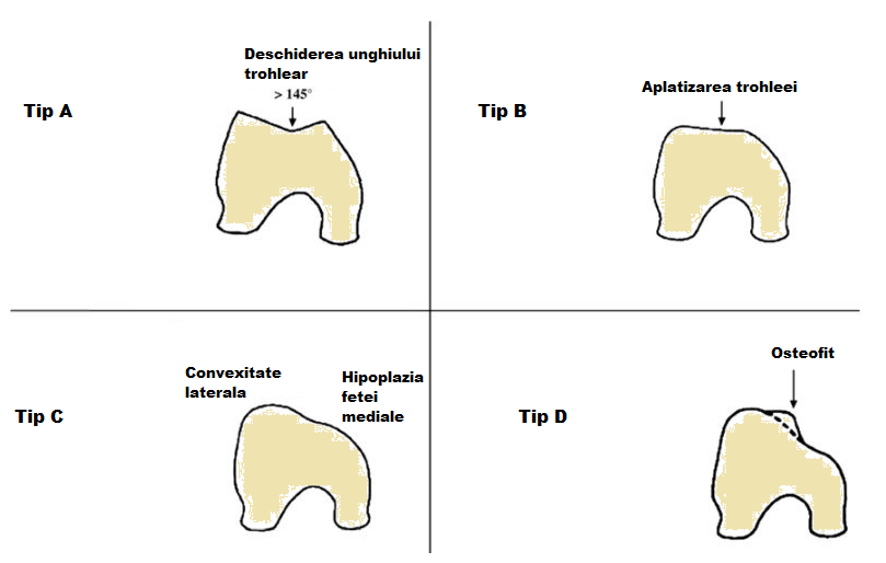
Fig. 21. TROCHLEAR DYSPLASIA - Dejour grading
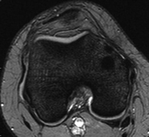
Fig. 22. A. MRI IMAGING REPRESENTATION OF TROCHLEAR DYSPLASIA TYPES (Dejour A)
Trochlear morphology is preserved, but the trochlear groove is slightly shallow.
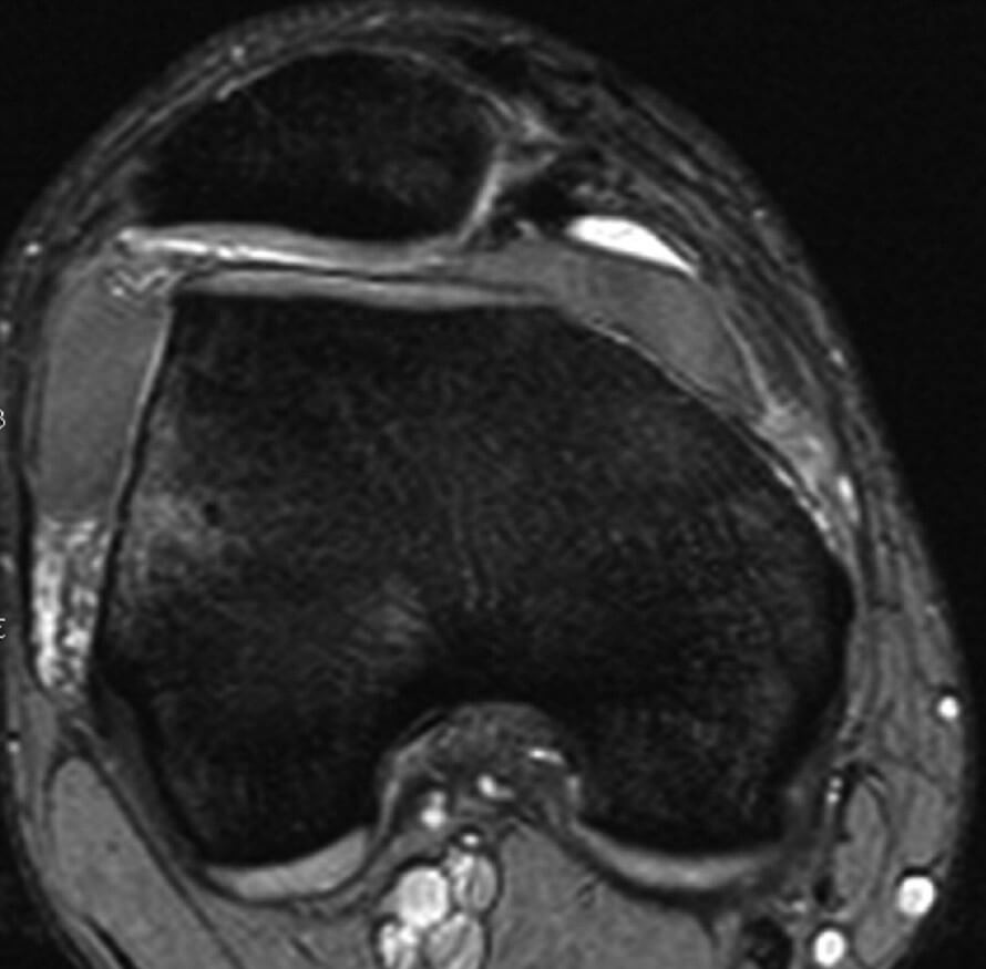
Fig. 22. B. MRI IMAGING REPRESENTATION OF TROCHLEAR DYSPLASIA TYPES (Dejour B)
Flat, horizontal surface of the trochlear joint.
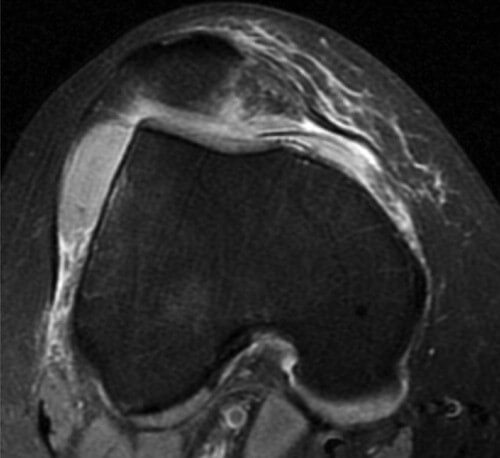
Fig. 22. C. MRI IMAGING REPRESENTATION OF TROCHLEAR DYSPLASIA TYPES (Dejour C)
Flat, obliquely oriented articular surface, with facet asymmetry.
Patella alta, or high-riding patella, is a patella that is situated above the trochlear fossa and occurs when the patellar tendon is too long. Patella alta is considered a major factor in patellofemoral malalignment because a greater degree of flexion is needed for the patella to engage in the trochlea compared with a normal knee. This problem leads to a reduction in the patellofemoral contact area and decreased bony stability at small degrees of flexion. Also, a true genu varum deformity of the knee contributes to a high patellar position by shortening the distance between the myotendinous junction of the quadriceps and the tibial tuberosity. In this situation, the extensor mechanism becomes the hypotenuse of a triangle, and this configuration can predispose to lateral patellar subluxation.
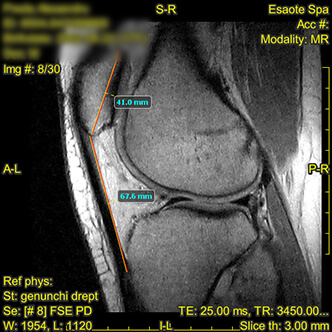
Fig. 24. SAGITTAL MRI SECTION IMAGE - FSE PD SHOWING A HIGH-RIDING PATELLA
The patellar height ratio evaluated on sagittal MR images is called the Insall-Salvati index. The length of the patellar tendon is measured from the patellar tip to its attachment at the tibial tuberosity and is divided by the longest superoinferior diameter of the patella to obtain the patellar height ratio. A patellar height ratio greater than 1.3 indicates a high-riding patella (patella alta).
Injuries to the medial ligamentous stabilizers, such as the medial patellar retinaculum, are diagnosed on MR images in 70%-100% of patients examined after lateral patellar dislocation. MRI imaging has a sensitivity greater than 80% when correlated with the clinical exam. Dividing the retinaculum into two or three regions appears to be useful for reporting the location of the injury: (a) the patellar insertion (anterior third), (b) the middle segment, and (c) the femoral origin (posterior third). Between 50% and 90% of injuries involve the patellar insertion. About a quarter of these patients have additional damage to a second region, and a fifth of patients present an injury in all three portions. In a quarter of cases, the MPFL is torn at the femoral insertion, which may also be accompanied by an avulsion injury of the femoral epicondyle. Precise characterization of the type of ligament injury is important, since results from the most recent studies indicate that femoral avulsion is a predictor of chronic instability, which is why the location of the injury can directly influence surgical planning.
A complete injury of the medial retinaculum is seen on MRI as a complete disruption of the ligament with the presence of edema in the adjacent soft tissues, in T2 hyperintensity. A lax or retracted fibrillar appearance with periligamentous edema indicates a complete disruption.
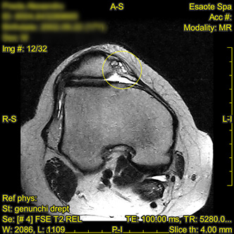
Fig. 25. PATELLAR RETINACULUM INJURY, ANTERIOR PATELLAR INSERTIONAL THIRD
The femoral trochlear groove shows hypoplasia of the medial facet with widening of the trochlear angle, an appearance corresponding to Dejour B trochlear dysplasia. The patella in turn shows a compensatory modified morphology, Wiberg type III.
The medial patellar retinaculum shows increased thickness, with a lax appearance, partially disorganized fibrillar structure, and intra- and periligamentous signal changes at its patellar insertion, with a small avulsed bone fragment of the medial patellar aspect. The MRI findings are suggestive of an injury to the anterior 1/3 of the medial retinaculum.
Lateral retinaculum with preserved course and signal.
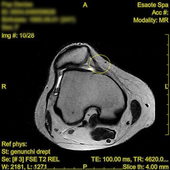
Fig. 26. MIDDLE 1/3 INJURY OF THE MEDIAL PATELLAR RETINACULUM
The medial retinaculum has a morphologically altered character, focally thickened in its middle segment, an appearance suggestive of a chronic injury. We note lateral patellar subluxation with chronic instability.
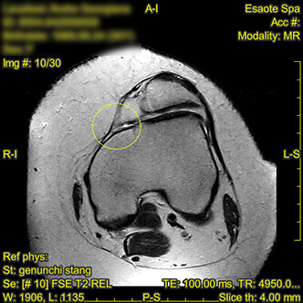
Fig. 27. INJURY OF THE MEDIAL PATELLOFEMORAL LIGAMENT - POSTERIOR 1/3
Injury detected at the posterior 1/3 condylar insertional level of the medial patellofemoral ligament. We note the lax, partially disorganized appearance at this level. The trochlear groove presents a Dejour B dysplastic character. The patella shows a single articular facet.
 |  |
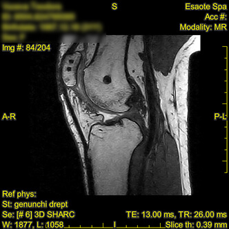 | 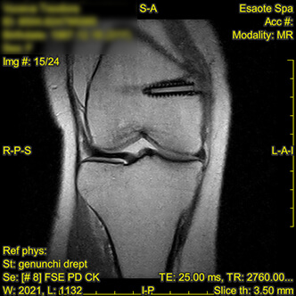 |
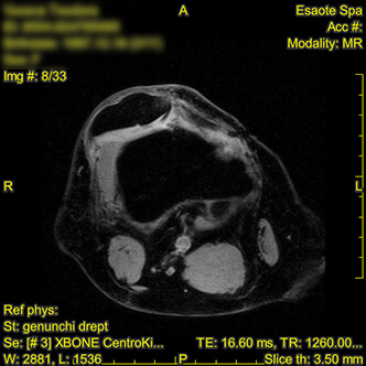
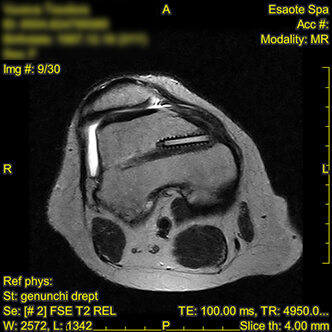
Fig. 28. RECONSTRUCTION OF THE MEDIAL PATELLAR RETINACULUM - MRI FINDINGS
The goal of surgical reconstruction is to restore the function of the medial stabilizers, which can be achieved by replacing a torn retinaculum with an autograft (gracilis tendon) or an allograft (for example, semimembranosus) (4). Various repair techniques have been described, but they all aim to stabilize the attachment of the retinaculum to the medial femoral condyle, its lowest point, or to attach it to the medial patellar facet. Approaches use different anchoring techniques and graft material. Mechanical stability often exceeds that of an intact native retinaculum, which is necessary to compensate for additional abnormalities that contribute to instability and predispose patients to future patellar subluxations or dislocations.
Centrokinetic is the place where you will find answers and clear solutions for your mobility problems. The clinic dedicated to bone and joint conditions is divided into the following specialized departments:
Find out the latest news by following the Facebook and YouTube accounts of the Centrokinetic clinic.
See details
READ MORESee details
READ MORESee details
READ MORESee here how you can make an appointment and the location of our clinics.








CENTROKINETIC FLOREASCA
Bucharest, 1st District,
Mircea Eliade Street, no. 18,
Eliade Tower, Entrance A, Floor 3
| Navigation | Explore 3D |
CENTROKINETIC Barbu Vacarescu
Bucharest, 2nd District,
Barbu Vacarescu Street, no. 102,
102 The Address, ground floor.
| Navigation | Explore 3D |
CENTROKINETIC MILITARI
Bucuresti, Sector 6,
Bld. Iuliu Maniu, nr. 15G,
Cladirea PBT, etaj 3.
| Navigation |
CENTROKINETIC CLUJ-NAPOCA
Cluj-Napoca,
Str. Intre Lacuri, nr.1
Intrarea este in spatele cladirii.
| Navigation |
SOCRATES CLINIC TIMISOARA
Timisoara, Calea Martirilor 1989,
nr. 1.
Telefon: 0371 785 374.
| Navigation |
Phone: 0319693
E-mail: programari@centrokinetic.ro
Operating hours:
Monday – Friday: 07:00 – 21:00
Saturday: 09:00 – 14:00
Sunday: closed.
MAKE AN APPOINTMENT