See details
READ MORE

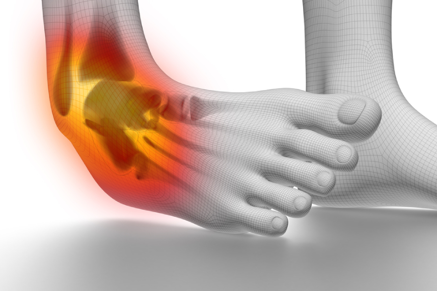
Joints need the stability provided by ligaments, bone structure, and muscles. If a joint is forced, a sprain can occur, meaning that the ligaments (bands of fibrous tissue that allow joint mobility) are stretched too much, or are partially or completely torn.
A sprain is an injury to a joint, caused by a sudden and forceful movement in which the ligaments stretch too much or tear. Sprains can be caused during household, sports or traffic accidents. Sprains most commonly occur in young, active, male adults. In terms of frequency, the most common are ankle sprains, followed by knee sprains, wrist sprains and thumb sprains.

Factors favoring sprains are:
Watch the video in which Dr. Gican Catalin, senior orthopedics-traumatology physician, talks to us about sprains:
In this article we explain what a sprain is, how many types of sprains are known, and what the symptoms and treatment options are for this injury.
.png)
A sprain is a stretching or tearing of the ligaments. A sprain can occur at any joint, but the most affected are the ankle, knee and wrist. Treatment for a sprain initially includes rest, ice compresses, compression and elevation. Mild sprains are treated very well at home. Severe sprains sometimes require surgery to repair the affected ligaments.
A sprain is different from a dislocation, as well as from a muscle strain. A sprain involves the displacement of a bony extremity (in most cases due to an osteoarticular trauma), while a muscle strain is an injury to the muscle fibers. Joint injuries occur either during a sudden movement or during physical activity, because that is when the ligaments are stressed beyond what their elasticity allows.
The degrees of a sprain range from a small stretch to a complete tear of the ligaments, depending on the severity of the injury. In the specialized literature, sprains are classified into three types, depending on the severity of the ligament injury (mild, moderate and severe). Treatment for a sprain and recovery time are determined based on the severity of the injury.
A grade I sprain reflects mild stretching of the ligaments, with a low level of damage to the constituent fibers. This type of sprain is very common, and treatment poses no particular problems. There may be mild tenderness in the affected area, swelling is minimal, there is microscopic tearing of some fibers, and bruising is less common in grade one sprains.
A grade II sprain is considered moderate and involves an incomplete tear of a ligament, meaning that although a partial tear has occurred, not all the ligaments are affected. A grade II sprain manifests through moderate tenderness and swelling and is usually associated with limited movement. The patient's symptoms include moderate pain, tenderness and swelling (which frequently appear a few days after the injury). Physical examination of the patient indicates abnormal weakness at the affected joint. Bearing weight can most often be difficult and painful. Medications such as acetaminophen, ibuprofen or aspirin can relieve pain. Also, rest, applying ice compresses and elevating the leg help reduce swelling. The patient requires physical therapy to more easily restore joint function. Typical recovery time for a grade II sprain is one to two months.
Grade III sprain represents a complete tear of the ligaments. This type of sprain is the most serious because it tears the ligament and in the most severe cases affects some smaller bones. This type of injury leads to greater instability of the joint involved. A grade III sprain can occur at any joint, but the ankle, wrist, knee and finger joints are most commonly affected. In the case of this sprain, the joint has significant instability, the area is very tender, and swelling, pain and bruising occur. Bearing weight causes severe pain.
Treatment for a severe sprain includes immobilizing the joint with a splint or a cast. The patient requires an extensive physical therapy and rehabilitation program, carried out over a period of several months, to facilitate the restoration of joint function. Grade III ankle sprains can lead to permanent, residual instability of the joint, increasing the risk of repeated ankle injuries. Severe sprains that do not respond to immobilization and physical therapy may require surgical reconstruction of the torn ligament, if the orthopedic doctor recommends it.

Sprains occur when the body is subjected to physical stress; in these situations, the muscles and joints must perform movements (sometimes sudden) for which they are not prepared or designed. A sprain can occur following a single stressful incident (in the case of contact sports, for example), or following several repetitions of a sudden movement. Typically, the injury mechanism involves excessive stretching of a tendon or ligament, which leads to its damage.
A sprain occurs when a ligament is stretched excessively or forced, in this way the joint is subjected to severe stress. Most sprains occur at the:
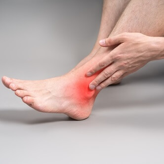
The symptoms of a sprain are varied and include bruising, pain, swelling and limited movement in the affected joint. A sprain can also lead to long-term health problems. For example, a wrist sprain can lead to the development of carpal tunnel syndrome. Symptoms can progress to numbness, tingling and pain, occurring during the day.
Another important symptom of a sprain is the inability to bear weight on the affected leg (a sprain can affect the ankle or the sole of the foot), due to pain and joint instability. This is more common in grade II and III sprains.
During the physical exam, the orthopedic doctor checks for swelling and tender points on the affected limb. The location and intensity of the pain help the doctor determine the extent and nature of the damage. X-rays can help rule out a diagnosis of fracture or other bone injuries. Also, magnetic resonance imaging (MRI) can be used to help determine the degree of the injury.
Centrokinetic benefits from state-of-the-art equipment and technology for investigations. If the sprain is severe, the doctor may recommend one or more imaging scans, to rule out the possibility of a broken bone, or to more thoroughly assess the extent of the ligament injury:
The orthopedic doctor will perform a physical exam to determine which ligaments have been affected (torn). During the consultation, the orthopedic doctor will move the joint in various directions, to check its mobility. To diagnose a sprain, the doctor will gently palpate the painful area.
Range of motion and resistance tests around the injured joint may also be performed. Many special tests, such as the anterior drawer test for the ACL at the knee, or the anterior drawer test at the ankle, are based on pulling the joint to test for excessive mobility. These tests give the doctor clues as to whether a ligament sprain may be present. An MRI is usually needed to determine whether a sprain is grade I, II or III.

Mild sprains can normally be treated at home, using the RICE method: rest, ice, compression, elevate. In the case of more severe sprains, immobilization of the injured limb may be recommended, so the injury has time to heal. Severe sprains may require surgery on the tendon or ligament. Moderate to severe pain should be treated under a doctor's supervision, who may recommend painkillers and anti-inflammatories, available at pharmacies without a prescription.
The injured limb needs rest for recovery; any activity involving the injured joint must be stopped. In the case of the lower limbs, walking or standing should be avoided. Ice reduces swelling by cooling the injured area and reduces bleeding by causing constriction in the vessels. Use compresses to wrap the injured part, to reduce swelling.
Elevation involves raising the affected limb, to the level of the heart or higher. This helps reduce blood flow to the affected area and, therefore, reduce swelling.
Treatment for mild sprains (treatment for an ankle sprain, for example) includes rest and temporarily giving up sports or recreational activities that could affect the ankle. Applying ice packs and raising the foot helps a lot to reduce inflammation. The doctor may recommend using an elastic band, to support the ankle. Prescribed exercises that help with joint flexibility and endurance facilitate healing.
Another important piece of information concerns the question of how long it takes for a sprain to heal. Grade I ankle sprains heal in about four to six weeks. A physical therapist can help the patient maximize the stability and endurance of the joint, or the injured limb. The orthopedic-traumatology doctor may also suggest immobilizing the limb with a splint. In the case of a severe sprain (with a torn ligament), surgery may even be considered.
Analgesics and nonsteroidal anti-inflammatory drugs are frequently used to control pain and inflammation in the case of sprains. The orthopedic doctor is the one who determines the appropriate treatment for their patient. Pain control is the initial goal of treatment for a foot sprain or a hand sprain.
In most cases, over-the-counter medications useful for eliminating pain, such as ibuprofen (Advil, Motrin IB, others), naproxen (Aleve, others) or acetaminophen (Tylenol, others), are enough to manage the pain caused by a sprain. The doctor may also indicate the use of an anti-inflammatory, or an ointment for the sprain.
Immobilization of the affected limb followed by long-term physiotherapy is the general treatment for grade III sprains, but in severe cases the doctor may recommend surgical reconstruction, since conservative treatment is associated with long-term chronic pain and joint instability.
This type of surgery is usually performed with a one-day hospital stay. The patient is given a general anesthetic, along with an injection around the affected joint, so that they do not feel numbness and pain after the surgery.
In some cases, the surgeon first performs arthroscopy of the ankle or knee, to inspect the joint before repairing the ligament. The procedure itself involves an incision (cut) on the outside of the ankle, to allow the surgeon to locate the scar tissue in the torn ligament near the fibula bone and to perform a repair with sutures at the bone.
Immediately after surgery:
The specialist doctor is the one who recommends the physiotherapist who can provide the patient with a personalized rehabilitation program. Following this program, the patient will be able to walk normally, and the swelling will noticeably decrease. The physiotherapist is an important part of the recovery program. Their skills in explaining and monitoring the necessary exercises (flexibility and mobility) allow the patient to recover after surgery in a controlled manner. The physiotherapist recommends performing each exercise slowly; pain is an important guide in performing these exercises. The patient is advised to stop the exercise if they feel more than slight discomfort. The time and type of recovery exercises recommended for you may vary, depending on the recommendations of the doctor or therapist.
Benefits of physical exercise after surgery:
Regular stretching and strengthening exercises, performed before sports, fitness or work activities, help reduce the risk of sprains. If you have an activity that involves physical stress, regular exercise can help prevent injuries (such as sprains) from occurring. Warm-up exercises (such as light jogging) before an intense activity can help avoid sprains. Also, avoid running, exercising or playing a sport when you feel tired or feel pain.
You can protect your joints in the long term by working to strengthen the muscles around the injured joint. Consult the orthopedic doctor and find out which exercises are appropriate for stretching and stability. Also, use footwear that offers support and protection.
Find out the latest news about us by following the Facebook and YouTube accounts of the Centrokinetic clinic.
Sprains can fully recover if treated appropriately and in a timely manner. It is important to take measures to avoid recurrence. Prevention is important in the case of sprains; treatment must be followed in accordance with the orthopedic doctor's recommendations, in order not to risk complications or recurrences of the sprain.
At Centrokinetic, the best doctors and therapists are available for any joint or musculoskeletal problem, from the initial consultation and complete investigations within the clinic, to treatment and complete medical rehabilitation for spinal, traumatic or post-surgical conditions.
Centrokinetic is the place where you will find clear answers and effective solutions for musculoskeletal conditions. Our clinic, dedicated to musculoskeletal disorders, is organized into the following specialized departments: Stay up to date by following Centrokinetic on Facebook, Instagram, and YouTube.
See details
READ MORESee details
READ MORESee details
READ MORESee here how you can make an appointment and the location of our clinics.








CENTROKINETIC FLOREASCA
Bucharest, 1st District,
Mircea Eliade Street, no. 18,
Eliade Tower, Entrance A, Floor 3
| Navigation | Explore 3D |
CENTROKINETIC Barbu Vacarescu
Bucharest, 2nd District,
Barbu Vacarescu Street, no. 102,
102 The Address, ground floor.
| Navigation | Explore 3D |
CENTROKINETIC MILITARI
Bucuresti, Sector 6,
Bld. Iuliu Maniu, nr. 15G,
Cladirea PBT, etaj 3.
| Navigation |
CENTROKINETIC CLUJ-NAPOCA
Cluj-Napoca,
Str. Intre Lacuri, nr.1
Intrarea este in spatele cladirii.
| Navigation |
SOCRATES CLINIC TIMISOARA
Timisoara, Calea Martirilor 1989,
nr. 1.
Telefon: 0371 785 374.
| Navigation |
Phone: 0319693
E-mail: programari@centrokinetic.ro
Operating hours:
Monday – Friday: 07:00 – 21:00
Saturday: 09:00 – 14:00
Sunday: closed.
MAKE AN APPOINTMENT