See details
READ MORE

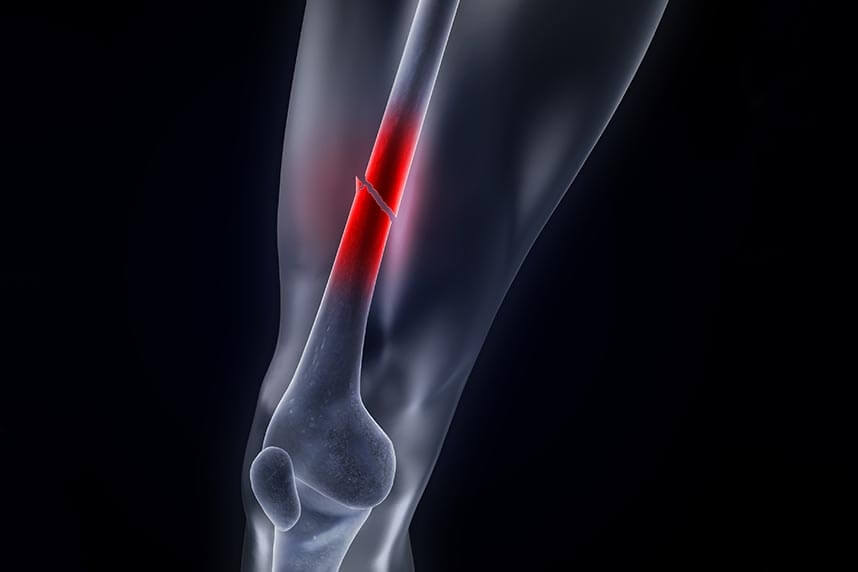
The femur, also known as the thigh bone, is one of the largest and strongest bones in the body, extending from the hip joint to the knee joint. Because it is so strong, significant force is required to fracture this bone. A femur fracture is a break in the thigh bone, or femur. The femur can be fractured in 3 areas: the head/neck of the bone (the upper end, near the pelvis), the shaft, or the lower end, near the knee. These injuries occur when a major-force impact strikes the thigh bone. This can be caused either by the person's own body weight (for example, a fall), or by a collision with an object (a car accident).
When the femur breaks, it takes a long time to heal. A fractured femur can make daily tasks difficult, since it is one of the main bones used for walking.
Femur fractures that are simple, short cracks in the bone, do not usually require surgery. However, fractures that break the bone completely, or that cause the bone to be displaced or crushed, usually require immediate surgery.
The type of fracture depends on where the forces come from. If there is a significant perpendicular force, this can lead to a transverse fracture. An axial force can travel upward and cause a fracture at the knee or hip. Rotational forces cause spiral or oblique fractures. The higher the energy, the higher the incidence of fragmented (comminuted) fractures. A femoral neck fracture occurs a few centimeters from the hip joint.
Femur fractures can be divided into three main types:
Proximal femoral fracture - A proximal femoral fracture, which occurs above the lesser trochanter, is generally referred to as a hip fracture.
Femoral shaft fracture - If the femur is broken between the proximal end and the distal end (where it meets the knee), then this is referred to as a femoral shaft fracture. This is a serious injury, generally associated with severe trauma, and in most cases requires surgery.
Supracondylar fracture of the femur - a rare injury, a supracondylar femoral fracture is a break in the bone near the knee joint. This injury generally damages the cartilage of the knee. A patient who suffers significant cartilage damage will very likely develop arthritis as they age.
Proximal femoral fractures, or hip fractures, involve the upper part of the thigh bone, adjacent to the hip joint. These fractures are further subdivided into different types:
Femoral head fractures are those that occur when the ball of the joint is broken at the top of the femur.
Intertrochanteric hip fractures occur just below the femoral head and are easier to repair than femoral shaft fractures.
Pelvic insufficiency fractures occur in the pelvic bone rather than the femur, and can often be treated without surgery.
A supracondylar fracture of the femur is an unusual injury that occurs just above the knee joint. These fractures often involve the cartilage surface of the knee joint, and are most commonly seen in people with severe osteoporosis or in those who have previously undergone total knee replacement surgery. A supracondylar fracture of the femur is a problematic condition, since it can increase the risk of knee arthritis later in life.
Early stabilization of the fracture is of paramount importance. It restores limb alignment, eliminates gross movement at the fracture site, limits further soft tissue damage, and reduces the risk of further spread of bacteria.
A femur fracture is generally the result of a strong blow. If the femur is compromised for some reason, such a fracture can be caused by milder injuries. The following are the most common causes of femur fractures:
Strong impact - In healthy people, generally a car accident or another extremely traumatic incident, a fall from a great height, or a gunshot to the femur can fracture the femur.
A fall - For elderly people who suffer from osteoporosis and a body generally weakened by age, and for people whose bones have been weakened by medication and/or conditions such as cancer, simply falling may be enough to suffer a femur fracture.
A pre-existing bone disease that weakens the bone, such as a tumor, Paget's disease, bone cysts, or osteoporosis.
There are several risk factors that can lead to a femur fracture. These include:
The different types of femur fractures have different symptoms. Common symptoms of a fractured femur:
If you have suffered a proximal femoral fracture:
If you have suffered a femoral shaft fracture:
If you have suffered a supracondylar femur fracture:
The symptoms of femur fractures can be confused with the symptoms of other medical conditions (for example, a patella fracture or a dislocated fibular head). Be sure to consult a doctor to find out whether you have a femur fracture, and get the appropriate treatment. For a correct diagnosis and a complete treatment plan, call on Centrokinetic! Our clinic has doctors highly specialized in orthopedics and a department dedicated to recovery.
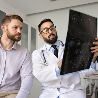
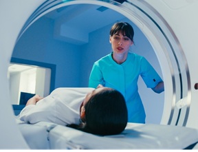
In most cases, your doctor will start with an X-ray. If more information is needed, the doctor may also perform a CT scan (computed tomography). Before recommending a specific treatment, your doctor will determine what type of fracture you have.
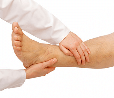
A femur fracture is a serious injury that is diagnosed by a doctor, usually in an emergency room. The doctor will check all the signs and symptoms of a fracture. The doctor will ask questions related to the location of the pain, whether any trauma or accidents occurred, factors that reduce/increase the pain, the mechanism of injury, and the time of onset.
It is important for your doctor to know how you injured your leg. For example, if you were involved in a car accident, it helps the doctor to know what speed you were going, whether you were the driver or a passenger, whether you were wearing a seatbelt, and whether the airbags deployed. This information will help the doctor determine how you were injured and whether you might be injured elsewhere as well. It is also important for your doctor to know whether you have other conditions such as high blood pressure, diabetes, asthma, or allergies. Your doctor will also ask you about any medications you are taking.
X-rays - X-rays use low-level radiation to photograph the bones. X-rays can show whether the bones are broken or intact.
CT scan - A CT scan uses X-rays and a computer to create a cross-section of the bone and tissue. This test will show whether the fracture is a simple break or whether the bone is broken into several pieces.
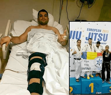
A femur fracture is always considered a medical emergency that requires immediate evaluation and treatment in a hospital. Treatment depends largely on the location of the fracture and the pattern and extent of the break.
Non-surgical treatment increases the risk of bone displacement and requires frequent X-ray monitoring to ensure proper healing. In more severe fractures, surgery is avoided only if the patient has serious medical conditions. Early treatment includes:
A femoral shaft fracture is a serious injury that usually occurs as a result of a high-speed car accident, or a fall from a great height. Treatment almost always requires surgery. The most common procedure involves inserting a metal rod (known as an intramedullary nail) into the center of the thigh bone. This helps reconnect the two ends, which are secured with screws above and below the fracture. The intramedullary nail generally remains in the bone but can be removed if it causes pain or other problems.
A less common technique involves the use of plates and screws to secure the fracture, which is then held in place by an external fixator. The fixator, which is located outside the leg but penetrates the skin to stabilize the bone segments, ensures that the femur is completely immobilized and able to heal better.
Patients with femur fractures begin rehabilitation through gradual, assisted physiotherapy, to restore muscle strength, hip flexibility, and prevent medical complications. This stage may require the patient to be admitted to a care center, or to receive regular home visits from a physiotherapist. During the first weeks up to 12 months after fracturing the femur, crutches or a cane may be necessary.
Physiotherapy in the first 6-12 weeks after surgery includes:
Complications that can occur with a femur fracture:
Correct alignment - If the femur is not aligned correctly, there is a chance that the injured leg will become shorter than the other one, and it can cause hip or knee pain many years later. Poor alignment of the femoral bone can also be painful.
Peripheral damage - The break can also injure the muscles, tendons, ligaments, and nerves of the leg.
Surgical complications - Some complications related to surgery include infections and blood clots.
A femur fracture can be prevented by avoiding the following risk factors:
In addition, strengthening the bones and muscles with weight-bearing exercises can also help reduce the risk of fracture.
In patients with osteoporosis, administration of the bisphosphonate group of medications is recommended, along with calcium and vitamin D supplements. Vitamin D is very important for strong bones, and a good source is the sun.
Physical exercise is not only essential for rehabilitation after a fracture, but also for strengthening bone mineral density, especially in vulnerable people, such as elderly patients, osteoporotic post-menopausal women, or people suffering from osteoporosis or osteopenia. Putting weight on the bone makes it provide more bone material to strengthen itself.
Bone density tests are recommended for all women aged 65 and older, and for younger women with an increased risk of fracture. Strong bones contain a dense framework of protein fibers covered with calcium. This support system decreases with age, lack of physical exercise, and low intake of calcium and vitamin D, among other reasons. Low bone density increases the risk of fracture.
Improving muscle strength is necessary to improve postoperative walking ability and to reduce the risk of falls. Physical exercise helps with:
Aerobic fitness is useful in a physical therapy plan, since improving cardiorespiratory capacity will lead to better walking ability.
Fall prevention programs are important for the elderly population who have already suffered a femoral fracture. The elderly often fall again after a previous hip or femur fracture, and this constitutes a major health problem. Prevention and treatment of fall risk factors are essential. These programs should include gait exercises and advice on assistive devices and medications, balance training exercise programs, treatments for cardiovascular problems, and environmental modifications.
Accident prevention: eliminating circumstances that cause injury - for example, reducing speed in traffic, wearing a seatbelt, installing support devices in the home, reducing alcohol consumption.
Centrokinetic is the most high-performing medical clinic dedicated to musculoskeletal conditions, organized into several dedicated departments:
Find out the latest news about us by following the Centrokinetic clinic's Facebook and YouTube accounts.
See details
READ MORESee details
READ MORESee details
READ MORESee here how you can make an appointment and the location of our clinics.








CENTROKINETIC FLOREASCA
Bucharest, 1st District,
Mircea Eliade Street, no. 18,
Eliade Tower, Entrance A, Floor 3
| Navigation | Explore 3D |
CENTROKINETIC Barbu Vacarescu
Bucharest, 2nd District,
Barbu Vacarescu Street, no. 102,
102 The Address, ground floor.
| Navigation | Explore 3D |
CENTROKINETIC MILITARI
Bucuresti, Sector 6,
Bld. Iuliu Maniu, nr. 15G,
Cladirea PBT, etaj 3.
| Navigation |
CENTROKINETIC CLUJ-NAPOCA
Cluj-Napoca,
Str. Intre Lacuri, nr.1
Intrarea este in spatele cladirii.
| Navigation |
SOCRATES CLINIC TIMISOARA
Timisoara, Calea Martirilor 1989,
nr. 1.
Telefon: 0371 785 374.
| Navigation |
Phone: 0319693
E-mail: programari@centrokinetic.ro
Operating hours:
Monday – Friday: 07:00 – 21:00
Saturday: 09:00 – 14:00
Sunday: closed.
MAKE AN APPOINTMENT