
Hernia, Lipoma, or Cyst? How Does a Soft Tissue Ultrasound Differentiate These Formations?


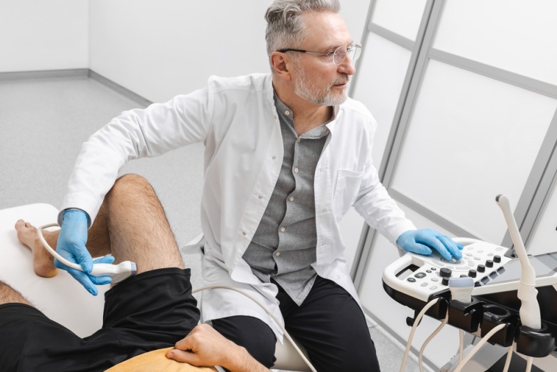 Hernia, Lipoma, or Cyst? How Does a Soft Tissue Ultrasound Differentiate These Formations?
Have you discovered a lump under your skin after an intense workout, after a more demanding period at the office, or simply by chance? Maybe it doesn't hurt, or maybe it only bothers you slightly, but you certainly want to find out what it is: is it a lipoma, a cyst, or a hernia?
For an active adult, an amateur athlete, or a parent attentive to their child's health, the difference matters. Each of these formations has different mechanisms, evolution, and treatments. Some require only monitoring, others have a surgical indication.
A soft tissue ultrasound helps you clarify the situation without exposure to radiation and without invasive investigations. With this investigation, the doctor can determine the nature of the formation and propose a personalized plan. Find out how these lesions differ and when it is time to book a consultation!
What is a lipoma: definition, characteristics, evolution
A lipoma is a benign tumor made of adipose (fat) tissue. It develops slowly, usually under the skin, and remains well delimited from the surrounding structures.
How do you recognize it?
In most cases, a lipoma:
- is soft to the touch;
- moves easily under the fingers;
- does not cause pain;
- grows slowly, over the years.
Lipomas frequently appear on the back, shoulders, arms, thighs, or abdomen. Most lipomas do not require treatment if they do not cause symptoms or aesthetic discomfort.
Advantages and limitations in evolution
Advantages (from a medical point of view):
- they are benign in the vast majority of cases;
- they grow slowly;
- they rarely cause complications.
Limitations or risks:
- they can compress a nerve and cause pain;
- they can grow beyond 5 cm, requiring additional investigations;
- very rarely, they can mask a malignant tumor (liposarcoma).
Certain atypical imaging characteristics (thick septa, increased vascularization) require additional imaging investigations to rule out malignancy. Do not try to treat a lipoma at home with ointments or improvised remedies. There is no solid scientific evidence to support their effectiveness!
What is a sebaceous (epidermoid) cyst: definition and particularities
The sebaceous cyst, correctly called an epidermoid cyst, is a subcutaneous cavity filled with keratinous material or semi-solid fluid. It forms through the blockage of a hair follicle or a sebaceous duct.
How do you recognize it?
Compared to a lipoma, a cyst:
- is firmer;
- can have a visible central point;
- can become red and painful if it gets infected;
- varies in size.
These cysts are frequently benign, but may require treatment if they become inflamed or recur.
Advantages and limitations
Advantages:
- they are benign lesions;
- they often remain stable.
Limitations:
- they can become infected;
- they can recur if the capsule is not completely excised;
- they can cause aesthetic discomfort.
Simple drainage removes the content, but if the doctor does not remove the capsule, the cyst can reappear.
What is a hernia: mechanism and major differences
A hernia is not a tumor; it represents the protrusion of an organ or tissue through a defect in the muscular wall.
The most common types are:
- inguinal hernia;
- umbilical hernia;
- femoral hernia;
- epigastric hernia.
How does it manifest?
A hernia has different characteristics compared to a lipoma or a cyst:
- it becomes more prominent with exertion or coughing;
- it can disappear when lying down (on your back);
- it can be reducible through gentle pressure;
- it can cause a feeling of heaviness or pain.
Untreated hernias can progress to incarceration or strangulation, situations that require prompt surgical intervention.
It is also important to differentiate the herniated disc, which affects the spine and produces radiating pain, numbness, or muscle weakness.
Why is palpation not enough?
The clinical examination provides clues, but does not always establish the final diagnosis.
A deep lipoma can seem fixed. An inflamed cyst can mimic a solid tumor. A small hernia can resemble an abdominal lipoma.
In addition, certain malignant soft tissue tumors can initially look similar to a lipoma. According to current data, a size over 5 cm, rapid growth, and inhomogeneous consistency require additional investigations. For stable and reliable results, you need imaging.
Soft tissue ultrasound: definition and benefits 
The soft tissue ultrasound uses ultrasound waves to visualize subcutaneous, muscular, and fascial tissues. It does not involve ionizing radiation and is suitable for adults, children, and pregnant women.
Advantages
- it is non-invasive;
- it allows real-time evaluation;
- it offers the possibility of dynamic examination (in motion, while coughing);
- it is accessible and fast.
Read more general information about ultrasound in our dedicated article. The experience of the imaging doctor influences the quality of the interpretation. A team specialized in musculoskeletal pathology correlates the images with the symptoms and establishes the appropriate course of action.
How does the ultrasound differentiate the lipoma, the cyst, and the hernia?
The lipoma on ultrasound
- homogeneous formation;
- well delimited;
- superficially located;
- without significant vascularization on Doppler.
If thick septa or inhomogeneous areas appear, the doctor may recommend an MRI for clarification.
The cyst on ultrasound
- anechoic or hypoechoic image (black appearance on the screen);
- fluid content;
- thin and regular walls;
- no internal vascular flow.
In case of inflammation, the wall can thicken, and the content becomes heterogeneous.
A particular type is the synovial cyst, frequently located at the joints of the hand or knee.
The hernia on ultrasound
- visible defect in the muscular fascia;
- protrusion of adipose or intestinal tissue;
- accentuation during the Valsalva maneuver;
- dynamic evaluation in real time.
The ultrasound measures the size of the defect and evaluates the content of the hernia sac.
Direct comparison between the three formations
This comparison helps you understand the differences, but does not replace a medical evaluation.
| Criterion | Lipoma | Epidermoid cyst | Hernia |
|---|---|---|---|
| Nature | Benign fatty tumor | Cavity with content | Muscular defect |
| Consistency | Soft, elastic | Firm or fluctuant | Variable |
| Mobility | Mobile | Semi-fixed | Changes with exertion |
| Pain | Rare | Possible with infection | Possible with exertion |
| Evolution | Slow | Variable | Progressive |
| Treatment | Monitoring/excision | Complete excision | Surgical |
When is an MRI or CT needed?
The ultrasound is the first recommended investigation. However, the doctor may indicate an MRI if:
- the lesion exceeds 5 cm;
- it is deep (intramuscular);
- it has atypical characteristics;
- malignancy is suspected.
The importance of early intervention
Intervention at the right time reduces the risk of complications. In the case of a hernia, scheduled treatment prevents incarceration. In the case of an inflamed cyst, early treatment reduces the risk of an abscess. In the case of a suspicious tumor, a rapid diagnosis allows the recommendation of an appropriate treatment plan, which may include, in some cases, procedures related to vascular surgery.
For athletes and active people, correctly identifying the problem allows a safe return to training.
Personalized approach and benefits for the patient
A correct evaluation offers you:
- diagnostic clarity;
- reduced anxiety;
- avoidance of unnecessary procedures;
- a treatment plan adapted to your age and activity level.
The medical team correlates the imaging results with the clinical examination and your history. For patients in post-operative recovery, ultrasound monitoring allows the physical therapy program to be adjusted.
Do not postpone the consultation if you notice changes in size, persistent pain, or skin changes. Prevention and regular check-ups support long-term health. Book an appointment now for a specialist consultation.
Frequently asked questions
Can the ultrasound reliably differentiate a lipoma from a liposarcoma?
In most cases, the ultrasound identifies the typical characteristics of a benign lipoma. If atypical signs appear, the doctor recommends an MRI or a biopsy for a definitive diagnosis. The combined evaluation increases accuracy.
Does a cyst need to be operated on immediately?
Not always. If it is small and asymptomatic, the doctor may recommend monitoring. If it becomes inflamed, painful, or recurs, complete excision is the appropriate option.
Does a small hernia require surgery?
Most hernias have a surgical indication, even if they are small, because the muscular defect does not close spontaneously in adults. The doctor determines the optimal timing of the procedure based on your symptoms and lifestyle.
Disclaimer: This article is for informational purposes and does not replace a specialist consultation.
Sources:
- "Lipoma: Usually Harmless Fatty Tumor-Lipoma - Symptoms & Causes - Mayo Clinic." Mayo Clinic, 2025. Accessed 8 Apr. 2026.
- Delgado, Amanda. "What's Causing This Cyst?" Healthline, Healthline Media, 15 Sept. 2015. Accessed 8 Apr. 2026.
- "Hernia: What It Is, Symptoms, Types, Causes & Treatment." Cleveland Clinic, 8 Sept. 2023. Accessed 8 Apr. 2026.
BUCHAREST TEAM


































































CLUJ NAPOCA TEAM


































BRASOV TEAM



















MAKE AN APPOINTMENT
FOR AN EXAMINATION
See here how you can make an appointment and the location of our clinics.
MAKE AN APPOINTMENT
Asiguratori privati





Parteneriate



CENTROKINETIC FLOREASCA
Bucharest, 1st District,
Mircea Eliade Street, no. 18,
Eliade Tower, Entrance A, Floor 3

| Navigation | Explore 3D |
CENTROKINETIC Barbu Vacarescu
Bucharest, 2nd District,
Barbu Vacarescu Street, no. 102,
102 The Address, ground floor.

| Navigation | Explore 3D |
CENTROKINETIC MILITARI
Bucuresti, Sector 6,
Bld. Iuliu Maniu, nr. 15G,
Cladirea PBT, etaj 3.

| Navigation |



CENTROKINETIC CLUJ-NAPOCA
Cluj-Napoca,
Str. Intre Lacuri, nr.1
Intrarea este in spatele cladirii.

| Navigation |


SOCRATES CLINIC TIMISOARA
Timisoara, Calea Martirilor 1989,
nr. 1.
Telefon: 0371 785 374.

| Navigation |
CONTACT
Phone: 0319693
E-mail: programari@centrokinetic.ro
Operating hours:
Monday – Friday: 07:00 – 21:00
Saturday: 09:00 – 14:00
Sunday: closed.
MAKE AN APPOINTMENT

