See details
READ MORE

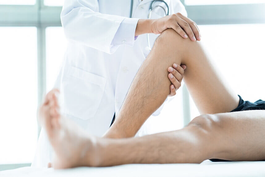
Estimating the final inequality at the end of growth
Estimating the final inequality at the end of growth is crucial for establishing treatment, and several examinations of the patient are necessary in order to achieve the greatest possible accuracy.
Treatment options
Inequalities smaller than 2 cm are most often asymptomatic; they are relatively common in the population, so only periodic monitoring is needed to detect any potential progression or complication.
A shoe lift should be considered when the inequality exceeds 2 cm and the patient walks on the tip of the shorter (equinus) foot.
An orthopedic boot is used in cases of shortening of 3-6 cm, as it better stabilizes the foot.
Temporary orthopedic compensation should be applied to patients awaiting surgery in order to avoid the occurrence of complications.
Epiphysiodesis: consists of ablation of the growth cartilage of the longer limb; it should be considered when the predicted final inequality is greater than 2 cm.
Surgical shortening of the longer limb is performed in patients with a mature skeleton with an inequality between 2-5 cm at the femur, or 2-3 cm at the tibia.
Limb lengthening by mechanical means can be performed acutely for a femoral shortening of 2-4 cm, or 2-3 cm on the tibia.
Combined techniques: shortening the longer segment by half the difference and acute lengthening of the short one.
Progressive lengthening with an external fixator, in inequalities exceeding 4 cm, the method of choice for patients who also have an axial deformity requiring correction.
Hybrid lengthening techniques with an external fixator combined with internal fixation using intramedullary rods, which have the advantage that the external fixator can be removed immediately after the distraction phase.
Expandable rods are the newest method for bone growth and consist of implanting intramedullary fixation devices capable of performing distraction (lengthening) using radio waves or electromagnetic field activation to control the rhythm and rate of lengthening. They have the advantage of a less invasive intervention – minimal postoperative scarring, lower risk of infection, and correction controlled by the doctor.
Contraindications
Lengthening: inequalities smaller than 3 cm, predicted shortening that cannot be compensated through lengthening, patients at risk of neurovascular injury or with poor bone quality, neurofibromatosis, irradiation.
Shortening: very young patients, patients presenting shortening >5% of the normal length (4 cm) in the apparently healthy limb, epiphysiodesis as the sole method for correcting inequalities greater than 8 cm; epiphysiodesis performed at too young an age, which will lead to a reverse inequality, or performed after the optimal time, which will fail to equalize the limbs.
External fixator: unstable joints above and below the segment to be lengthened, non-compliant patient, inadequate care (risk of infection).
What does Centrokinetic offer you if you have a leg length inequality?
• an accurate diagnosis through a clinical medical examination and imaging investigations
• a highly specialized team
• proper coordination, with children or adults able to benefit from consultations in neurology, orthopedics, and medical recovery.
Centrokinetic is the most high-performing medical clinic dedicated to osteoarticular conditions, organized into several dedicated departments:
Find out the latest news about us by following the Facebook and YouTube accounts of Centrokinetic clinic.
See details
READ MORESee details
READ MORESee details
READ MORESee here how you can make an appointment and the location of our clinics.








CENTROKINETIC FLOREASCA
Bucharest, 1st District,
Mircea Eliade Street, no. 18,
Eliade Tower, Entrance A, Floor 3
| Navigation | Explore 3D |
CENTROKINETIC Barbu Vacarescu
Bucharest, 2nd District,
Barbu Vacarescu Street, no. 102,
102 The Address, ground floor.
| Navigation | Explore 3D |
CENTROKINETIC MILITARI
Bucuresti, Sector 6,
Bld. Iuliu Maniu, nr. 15G,
Cladirea PBT, etaj 3.
| Navigation |
CENTROKINETIC CLUJ-NAPOCA
Cluj-Napoca,
Str. Intre Lacuri, nr.1
Intrarea este in spatele cladirii.
| Navigation |
SOCRATES CLINIC TIMISOARA
Timisoara, Calea Martirilor 1989,
nr. 1.
Telefon: 0371 785 374.
| Navigation |
Phone: 0319693
E-mail: programari@centrokinetic.ro
Operating hours:
Monday – Friday: 07:00 – 21:00
Saturday: 09:00 – 14:00
Sunday: closed.
MAKE AN APPOINTMENT