Actualizat: 17-03-2025 / Publicat: 14-03-2018
This article was written by
Dr. Crintea Alexandru, orthopedic specialist, with over 10 years of experience, further specialized through numerous national and international courses.
A meniscus injury is generally caused by a twisting motion between the femur and tibia without contact, or a sudden movement such as a squat. Any movement that allows the femur to slide too far forward or backward relative to the tibia can cause some of the forces to be transmitted to the meniscus and lead to a meniscus tear.
A meniscus tear is an injury that occurs when pieces of the meniscal cartilage are torn, due to specific movements that rotate the knee suddenly or forcefully, or due to other factors. Meniscus injuries are often seen in athletes as a result of a sports accident and account for approximately 15% of all sports injury cases. There are partial or complete tears of a lateral or medial meniscus. Medial tears are reported more often than lateral tears. In addition to tears that occur in sports, osteoarthritis can also lead to a spontaneous tear of the meniscus by weakening the meniscal structure. Possible risk factors for meniscal tears are sports, older age, male sex, and pre-existing conditions such as osteoarthritis.
Like many other knee injuries, a meniscus tear can be painful and debilitating. Unfortunately, it is quite common. In fact, a meniscal tear is one of the most common cartilage injuries in the knee.
So, what is the meniscus? It is a piece of cartilage in the knee that cushions and stabilizes the joint. It protects the bones from wear. But all it takes is one good twist of the knee to tear the meniscus. In some cases, a piece of the torn cartilage breaks off and gets caught in the knee joint, causing it to lock.
Meniscus tears are common in contact sports, such as football, as well as sports that require jumping, such as volleyball. Meniscus tears are a particular risk for older athletes, since the meniscus weakens as we age. More than 40% of people aged 65 or older suffer from meniscus tears.
![recuperare ruptura de menisc extensii spalier]()
A meniscus fissure is a partial tear of the meniscus. It can occur with a sudden movement of the thigh while the foot remains planted. Both the inner and outer meniscus can be injured during sustained effort, which is why this problem occurs especially in performance athletes.
The main symptoms of a meniscus fissure are similar to the symptoms of a meniscus tear:
- Knee locking
- Stiffness and swelling of the knee
- A feeling of knee instability
The meniscus can be torn during activities that cause direct contact or pressure from a forced twist or rotation. A sudden pivot or turn, squats, or heavy lifting can lead to injury. Many athletes are at risk of tearing their meniscus.
Sports that require sudden turns and stops can put you at greater risk for a meniscus tear. Some of these sports include:
- football
- basketball
- rugby
- tennis
Meniscus tears also appear to be becoming more common in children. This is because they are participating in organized sports at a younger age. In addition, when a child focuses on a single sport, they are more likely to experience a meniscus tear. The same is true for teenagers who participate in competitive sports.
The meniscus weakens with age. Tears are more common in people over the age of 30. Movements such as squatting or standing up can lead to injury in a person with a weakened meniscus.
If you have osteoarthritis, you are at a higher risk of injuring your knee or tearing your meniscus. Osteoarthritis is a common condition that involves pain and stiffness in the joints caused by aging and wear. When an older person has a meniscus tear, it is more likely that the cause is related to degeneration. This happens when the cartilage in the knee becomes weaker and thinner. As a result, it is more prone to tearing.
![fizioterapie genunchi]()
Meniscus tears are very widespread. Depending on your age, there are different types of meniscus tears that could be present in the knee. Not all meniscus tears are the same. They were not all caused by the same trauma. And most importantly of all, not all meniscus tears should be treated the same way. Having knowledge about the different types of meniscus tears will help you understand what treatment, if any, can improve your knee pain.
First, there are acute or traumatic meniscus tears, which occur as a result of an injury. Then, there are degenerative-type meniscus tears that occur as a result of wear and tear, and are usually not preceded by trauma. Traumatic meniscus tears come in all shapes and sizes. There are radial tears, short tears that extend from the inner edge of the meniscus and head toward the periphery or outer part of the meniscus. The largest of all meniscus tears is a bucket-handle tear, which is "c"-shaped and looks like a bucket handle.
A knee examination and possible arthroscopy provide a clear classification of meniscus injuries. The size and severity of the meniscus tear are often indicative of recovery after meniscus surgery and any subsequent degenerative phenomena in the knee joint.
Different types of meniscus tears:
- Radial tear
- Oblique tear
- Longitudinal tear
- Bucket-handle tear
- Horizontal tear
- Complex tear
![exercitii recuperare ruptura de menisc bicicleta medicinala]()
When a meniscus tear occurs, you may hear a sound around the knee joint. After that, you may experience:
- pain, especially when the area is touched
- swelling
- difficulty moving the knee or an inability to move it through a full range of motion
- a feeling of the knee locking or catching
- a feeling that the knee is giving way or not offering support
You may also feel a slipping or popping sensation, which is usually an indication that a piece of cartilage has become loose and is blocking the knee joint.
Knee pain
Knee pain is very common. It can appear out of nowhere, after a tennis match, a long walk, or an accident. One of the most common reasons for knee pain is a meniscus tear. In many cases, pain from a torn meniscus will improve significantly or disappear without surgery. In cases where there was an injury that preceded the meniscus tear, you may have knee pain and swelling in almost all daily activities. These acute meniscus tears usually occur in active young people. The tear can cause mechanical pain, such as popping, locking, or even causing the knee to catch or become unstable. Most people with acute, traumatic meniscus tears should consider surgery to repair the tear.
Knee swelling and stiffness
Knee swelling can limit knee flexibility and function. For example, a person with a swollen, stiff knee will find it difficult to fully bend or fully straighten the knee, and the joint may naturally bend 15 to 25 degrees while the leg is at rest.
Depending on the underlying condition, a swollen knee may present no other symptoms, or it may be painful, red, and difficult to put weight on.
Knee locking
The most common symptoms of a meniscus tear are knee pain, swelling, stiffness, instability, and sometimes locking - where the knee becomes stuck in a single position.
"Knee locking" or "locked knee" is the term used to describe a painful condition that occurs as a result of extending the knee to a certain angle. During extension of the knee joint, extension is limited to 10 to 30 degrees to prevent pain. The patient cannot reach an optimal normal extension. Any further extension beyond the limited angle causes severe knee pain. The most common cause of knee locking is a meniscus tear, congenital defect, injury, or diseases such as osteoarthritis.
Knee locking is a condition caused by inflammation of the synovial membrane, cartilage damage, ligament rupture, or a fracture of the patella, tibia, or fibula. The knee joint is a compact joint. The space within the knee joint is limited and narrow. Trauma, injury, and conditions that cause hematomas, mucosal edema, or inflammation of the synovial membrane in the joint can compromise the joint space and restrict extension of the knee joint.
Extension and flexion of the knee joint depend on the alignment of the medial and lateral meniscus, as well as the stability of the anterior and posterior ligament. The knee joint, during flexion and extension, maintains an optimal vertical, horizontal, or oblique position of the meniscus. Disorders of the meniscus or fragments in the knee joint interfere with achieving an optimal resting position in knee extension.
![recuperare genunchi bicicleta medicinala benzi kinesio]()
Hydrarthrosis
Excess fluid in the knee, or hydrarthrosis, occurs when excess fluid accumulates in or around the knee joint. There are many common causes of inflammation, including arthritis and ligament or meniscus injuries.
You might have hydrarthrosis if you notice:
- Swelling. The skin around the knee looks and feels puffy compared to the other knee.
- Stiffness. Range of motion decreases. The leg is difficult to fully bend or straighten. Depending on the cause, you may also notice knee instability or locking.
- Pain. This can range from mild to severe, depending on the cause.
- Swelling that appears in the absence of an injury may be caused by an infection. Additional symptoms include fever, chills, or redness.
Hemarthrosis
Hemarthrosis is blood inside the knee, or bleeding into the knee joint space. Fluid swelling inside the knee joint is usually reduced by aspiration. The needle is inserted from the side of the knee just above the upper edge of the patella. The needle enters under the patella into the suprapatellar bursa, which is continuous with the joint cavity.
If the aspirated fluid is not bloody, it is probably due to synovial irritation caused by chronic processes, such as gout, pseudogout, arthritis, rheumatoid arthritis, or a degenerative meniscus. A degenerated meniscus can be associated with swelling and fluid collection, which usually does not contain blood. The peripheral portion of the meniscus is vascular (approximately 3-5 mm). The blood supply to the meniscus comes from the medial and lateral genicular arteries. However, a traumatic meniscus tear can cause bleeding into the knee joint.
Knee instability
Knee instability is a condition characterized by a feeling of weakness in the knee, especially during movement. Some activities can place greater pressure on the knee joint and the structures surrounding it, which can cause instability in the knee. Although there are many causes of knee instability, knee joint injury is one of the most common causes.
The key to healing chronic knee pain is locating the points in the knee that are the cause of knee instability. Healing from knee pain requires finding the source of the knee pain, while also knowing and understanding the correct diagnosis.
Knee instability can be experienced when there is increased laxity of the ligaments that support the knee joint. It can be either physiological laxity or pathological laxity, in which an injury has caused the ligament to stretch.
Each meniscus is a C-shaped cushion, placed in each knee joint, located between the two bones that form the joint. They provide knee stability, support the knee, and act as a shock absorber, thus preventing damage to the knee joint.
Meniscus tears are another common cause of knee instability and can greatly affect knee function. Partial meniscus tears are more common and can cause knee instability, which does not cause discomfort but does cause even greater pain. This makes it difficult to move or turn the knee, put weight on it, or walk, and causes knee instability, which makes a person limp when trying to walk.
Other symptoms of a meniscus tear
Other symptoms of a meniscus tear will largely depend on the specific individual, as well as the location and degree of damage to the meniscus.
Symptoms may include:
- Pain localized near the tear area. In a lateral meniscus tear, this discomfort will be present along the outer edge of the knee. The pain will manifest on the inner edge of the injured knee.
- Immediate pain after the injury. A torn meniscus will often be evident from the moment the injury occurs. In these cases, the meniscus tear is usually accompanied by a popping sensation in the leg during an excessive twisting or stretching motion.
- Slow onset of symptoms. On the other hand, for some, the meniscus can tear without much initial sign or pain. This slow onset of symptoms is more common in older people and in those with knee cartilage damaged by osteoarthritis.
- Pain with movement. The pain will reflect the location of the tear, but will spread throughout the knee when it is in motion. If the knee locks, bending will cause the pain to worsen.
- Pain after rest. The pain will likely diminish with rest; however, in most cases it will return when you move. Movement can also worsen swelling.
- Fluid accumulation in the knee joint. This accumulated fluid will cause swelling of the entire area and reduced mobility. This symptom, which can occur as a result of a number of knee injuries, is known as "water on the knee."
- Knee locking. If a piece of the meniscus breaks off from the disc structure due to a tear, it can become lodged in the knee joint. This deposit can cause the knee to lock, in which a person loses the ability to fully straighten the leg while sitting or standing.
![extensie genunchi recuperare interventie ortopedica]()
Meniscus tears are common in sports, and in the general population as it ages. In acute injuries, such tears often occur with twisting movements while the knee is semi-bent and the foot is planted on the floor. Degenerative wear and tear of the meniscus, which tends to occur as we age, can develop with minor forces and daily activities. Large studies have analyzed the risk factors that correlate with meniscus tears. Such risk factors for degenerative tears include age over 60, being male, kneeling and squatting at work, and regularly climbing stairs. Prolonged walking and standing, carrying heavy loads, and being overweight are additional risk factors for such meniscal tears.
Acute meniscus tears have been found especially in football and rugby, and in other contact sports. People who have had ACL tears and gone at least 12 months without reconstructive surgery are also more likely to develop medial meniscal tears (on the inside of the knee), but not lateral meniscal tears (on the outside of the knee).
Complications following a torn meniscus can include knee instability, an inability to move the knee normally, or persistent knee pain. There is also a higher likelihood of developing osteoarthritis in the injured knee.
![recuperare genunchi spalier]()
The diagnostic process for meniscal tears aims to determine, first of all, whether a tear exists or whether the knee symptoms are caused by something else. From this point of view, diagnostic tests will try to determine which meniscus is torn, how severely it is damaged, and what treatment is recommended.
Patient history
The doctor will first assess whether a tear has occurred, by asking questions about the onset of symptoms. They will ask whether the pain was immediate, whether or not the pain was accompanied by a pop, and whether or not the knee was rotated.
The doctor will usually also consider and discuss the person's medical history before making treatment recommendations. For example, patients who have suffered multiple meniscus tears in the past may be more qualified candidates for a meniscectomy than those experiencing a meniscus tear for the first time.
Clinical examination
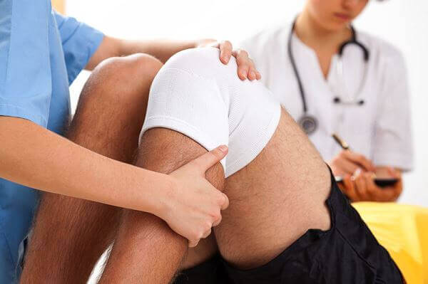
After determining the patient's medical history, the doctor will try to find out whether knee locking is present, whether there is a torn piece of cartilage, and whether the meniscus tear is lateral or medial. The doctor will assess the patient's range of motion by moving the leg in different directions.
The McMurray test is commonly used as follows:
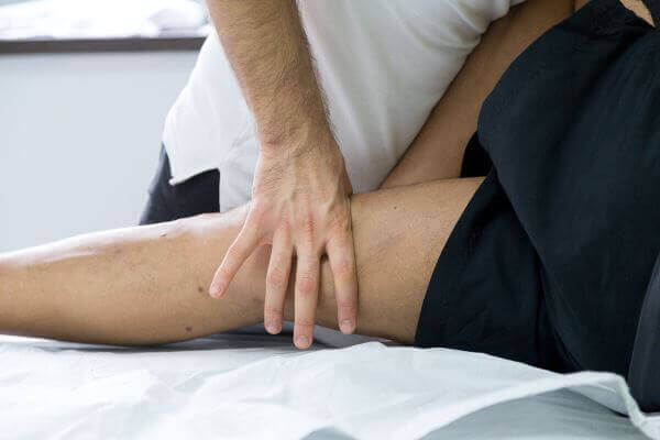
A doctor will flex the patient's leg to or beyond 90 degrees of extension, as if it were being straightened. Once the leg is straightened, the doctor will turn the leg in both directions, so that the tibia rotates over the femur both internally and externally. Internal rotation tests for a lateral meniscus tear, while external rotation tests for a medial meniscus tear. While this is being performed, the doctor lightly touches or palpates the knee.
The Apley compression test is commonly used as follows:
The patient lies on their stomach, bending the knee at a 90-degree angle. Downward force will be applied to the leg by the doctor while the leg is rotated outward. If pain presents in the inner section of the knee joint, a medial meniscal tear may be present.
Exploratory arthroscopy
Knee arthroscopy is a surgical procedure that allows doctors to see inside the knee joint without making a large incision through the skin and other soft tissues. Arthroscopy is used to diagnose and treat a wide range of knee problems.
During knee arthroscopy, the surgeon inserts a small camera, called an arthroscope, into the knee joint. The camera displays images on a video monitor, and the surgeon uses these images to guide miniature surgical instruments.
Arthroscopy can uncover several problems in the knee, such as a torn meniscus or an improperly aligned patella. Joint ligaments can also be repaired. There are limited risks to the procedure, and the outlook is good for most patients. The time and prognosis for recovery will depend on the severity of the knee problem and the complexity of the procedure required.
Imaging tests
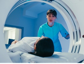
If surgery is being considered, a magnetic resonance imaging (MRI) scan of the knee may be ordered. The results of MRI examinations allow for a deeper view of the soft tissues in the knee joint, which can be factored into determining the location and severity of the tear. It will also indicate how much excess fluid is in the knee and whether or not a piece of meniscus has broken off inside the joint.
X-rays are useful in diagnosing degenerative arthritis in the knee joint, which could cause knee locking.
![recuperare dupa meniscectomie]()
The severity and location of the tear will be vital factors in determining a treatment regimen. For example, the outer third of the meniscus has a greater amount of blood supply than the inner tissue, and wounds there will heal faster, because blood brings nutrients that heal the injured tissue.
The patient's medical history, the individual's level of athleticism, and their desire to return to sports will be taken into consideration.
Treatment for a torn meniscus often begins conservatively, depending on the type, size, and location of the tear.
Tears associated with arthritis generally improve over time with arthritis treatment, so surgery is usually not indicated. Many other tears that are not associated with a blockage of knee movement will become less painful over time, so they also do not require surgery.
Your doctor may recommend:
- Rest. Avoid activities that worsen knee pain, especially any activity that makes you twist, rotate, or pivot the knee.
- Ice. Ice can reduce knee pain and swelling. Use a cold pack, a bag of frozen vegetables, or a towel filled with ice cubes for about 15 minutes, keeping your knee elevated. Do this every four to six hours on the first day, and then as often as needed.
- Medication. Pain relief supplements can also help ease knee pain.
Physical therapy exercises
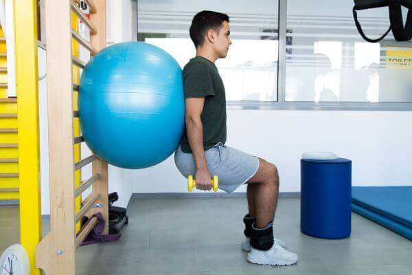
Exercise is an effective treatment for patients with knee degeneration, and aims to improve knee function and limit joint pain. There is strong evidence that physical therapy exercises play an important role in reducing symptoms, and improving muscle strength and physical capacity.
Important points in therapy:
- Controlling pain and inflammation
- Regaining control of the knee
- Regaining range of motion
- Restoring flexibility
- Restoring muscle function - The program must include both concentric and eccentric exercises in order to gain muscle hypertrophy as well as neuromuscular function.
- Optimizing neuromuscular coordination - proprioceptive re-education
- Progressive weight-bearing - weight-bearing exercises and reduced strain on the joints are necessary to improve functionality in meniscus repair, and should progress as indicated by the consultant. Excessive shear forces can be disruptive and should be avoided initially.
The RICE technique
If the tear is small and located on the outer edge of the meniscus, it may not require surgical repair. As long as symptoms do not persist and the knee is stable, non-operative treatment may be all you need.
The RICE protocol is effective for most sports-related injuries. RICE stands for rest, ice, compression, and elevation.
Rest. Take a break from the activity that caused the injury. Your doctor may recommend using crutches to avoid stress on the leg.
Compression. To prevent further swelling and blood loss, wear an elastic compression bandage.
Ice. Use cold packs for 20 minutes, several times a day. Do not apply ice directly to the skin.
Elevation. To reduce swelling, lie down when resting and place your leg on a pillow, above heart level.
Medication treatment
Non-steroidal anti-inflammatory drugs, such as Advil, Aleve, or Motrin, will help with pain and swelling. However, these medications can have side effects, such as an increased risk of bleeding and ulcers. They should only be used occasionally, unless your doctor expressly tells you otherwise.
Orthopedic treatment
Quite often, meniscus injuries can also be treated through non-operative interventions. In these cases, orthopedic treatments may include physical therapy techniques, exercise regimens, and/or anti-inflammatory and pain medications.
An elastic compression bandage can help in the short term to reduce swelling and provide some support to the knee. Long-term use is not recommended because it can lead to muscle weakening.
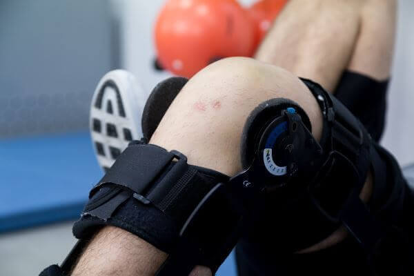
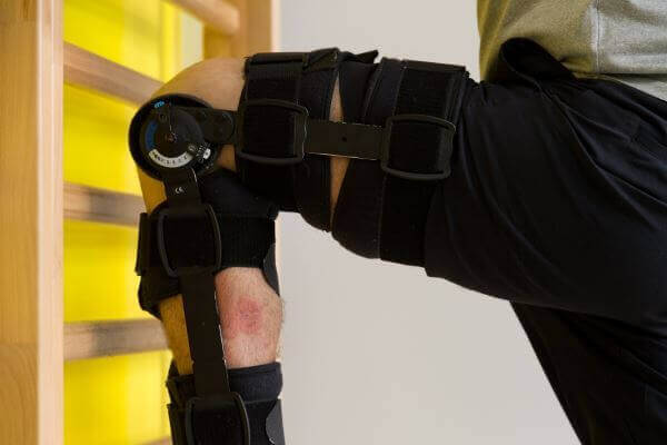
A knee brace offers much more stability than an elastic bandage, so it can be very useful, especially if the knee feels unstable. Usually, an advanced knee brace will provide sufficient support.
![examinare medicala genunchi ruptura de menisc]()
If conservative therapy fails, surgery may be considered. Knee arthroscopy allows the orthopedic surgeon to assess the cartilage tear and repair it. During an operation, the goal is to preserve as much cartilage as possible. Procedures include
meniscus repair (suturing the torn edges), partial
meniscectomy (cutting out the torn area and smoothing the injured site), or total meniscectomy, removing the entire meniscus, if this is considered appropriate.
Partial meniscectomy
Two small incisions are made in the front region of the knee below the patella. A camera is inserted through one of the incisions, so the surgeon can see inside the knee joint on a monitor. The other incision is used to place a tool in the joint that will grasp and remove the torn piece of cartilage. While the camera is inside the joint, the surgeon uses this opportunity to examine the rest of the knee. For most tears, the damaged portion of the meniscus will be removed and then sutures are used. This intervention is referred to as a partial meniscectomy. In a complete meniscectomy, the entire meniscus is removed, although this poses an increased risk for future complications. Studies on partial meniscectomy have shown that 78% to 88% of people have good results following this intervention. This means that 78 to 88 out of 100 people who have this surgery have reduced symptoms and can return to most or all of their activities.
Total meniscectomy
If a total meniscectomy is necessary, that means the entire meniscus is removed. Removing the entire meniscus generally reduces some symptoms. But losing the meniscus reduces the joint's cushioning and stability. Most people, especially if they are young or active, are not satisfied with a total meniscectomy. This is why surgeons try to remove as little of the meniscus as possible.
Meniscus transplant
If a meniscus is so severely damaged that it cannot be repaired, it may need to be removed or trimmed. Without the meniscus cushion, persistent knee pain and arthritis can develop. For many older patients who have this condition, knee replacement might be the right option. But active people under 55 may be eligible for an alternative treatment: meniscus transplant. A meniscal transplant replaces the damaged meniscus with donor cartilage. The goal of meniscus transplant is to replace the meniscus cushion before the joint cartilage becomes damaged. The donor cartilage supports and stabilizes the knee joint. This eases knee pain. The hope is that the transplant will also delay the development of arthritis, but long-term results are not yet available.
![terapie manuala genunchi]() Knee rehabilitation
Knee rehabilitation varies depending on the injury, the type of surgery, the orthopedic surgeon's preference, age, health status, and your activities. Time periods vary, but meniscus surgery is usually followed by a period of rest, walking, and selected exercises. Most people who have arthroscopic meniscectomy can bear weight one or two days after surgery, and can return to full activity within 2 to 4 weeks. Once a full, pain-free range of motion is possible, you can return to your previous level of activity.
The time needed to return to walking, driving, and more vigorous activities will depend on the type and extent of the surgery and your success in rehabilitation.
In contrast, rehabilitation for meniscal repairs or replacements is much more extensive and is based on the biomechanical function of both knees. In the first six weeks, there is no weight-bearing, other than full axial loading. Range of motion is limited to between 60 and 90 degrees of flexion for 6 weeks for lateral and medial meniscus repair. After the first 6 weeks, weight-bearing and range of motion increase according to the functional status of the knee.
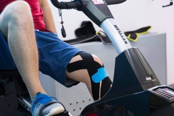
Rehabilitation treatment consists of ice therapy, ultrasound, friction massage, joint mobilization, cycling, etc. Underwater treatment cannot begin until the wounds have healed well, to prevent an increased risk of infection.
![fizioterapie genunchi]()
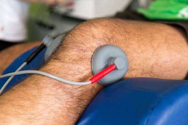
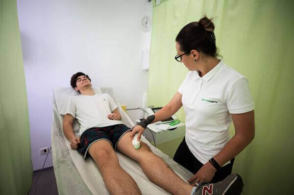
Neuromuscular electrical stimulation of the muscles around the knee can be used with the aim of strengthening the meniscus and surrounding tissues. Corticosteroid injections into the knee joint can be used to ease pain or inflammation in the soft tissue of the knee. Vibration therapy is another method applied by doctors. Extracorporeal shock wave therapy is also very useful. Gait therapy is another method that helps patients regain the normal posture they lose due to pain and discomfort.
Regenerative cell injections
Specific cell treatment for meniscus tears offers patients a minimally invasive injection procedure, which can help heal the injured tissue and allow the individual to avoid the painful, lengthy recovery that usually follows surgery, as well as the long-term impact on the knee, a result of removing sections of the meniscus.
These are cells from one's own body that can differentiate. They live in all of us in various tissues, ready to act to repair damage when it occurs. As we age or have large injuries, we may not be able to recruit enough cells to fully repair the area. Injection procedures help overcome this problem by extracting them from a high-volume area, then concentrating the cells and reinjecting them into the affected area to help the body heal naturally.
Injecting these cells has proven to be a better strategy for repairing degenerative cartilage than implanting differentiated cells such as articular cartilage. Adult cells have a reliable potential to differentiate into cartilage, bone, fat, or soft tissue. They also show the ability to cover areas of inflammation and degeneration and to modify the activity of the immune system, which can favorably influence the surrounding cartilage in the affected areas.
They can be extracted through minimally invasive methods from fat and from bone marrow. Cells from bone marrow have a good differentiation potential, and the concentrated cells have numerous growth factors that help with the cartilage-formation process. Unfortunately, the number of cells in bone marrow decreases dramatically as we age and they are often ineffective in elderly patients.
Cells derived from fat show an increased ability to differentiate into cartilage and are found in dramatically greater numbers than that observed in bone marrow. Not only are they typically 500-2000 times more abundant, but they do not decrease as we age. Fat-derived cells have been shown to have been used successfully to address cartilage defects and to improve osteoarthritis, in studies conducted on patients. Adult fat-derived cells have also demonstrated improved results over bone marrow for cartilage repair.
Cartilage growth factor injections
One component of blood flow is plasma. Platelets are cell-like particles that are smaller than red or white blood cells. These small platelets release growth factors at the site of injury. They help with the clotting process by sticking to the walls of damaged blood vessels. The most important compound is PDGF, or platelet-derived growth factor, which causes the release of specific cells from the vessel walls and their migration to the site of injury. Doctors can take a small amount of blood from a patient, isolate and concentrate the platelets by spinning the blood in a centrifuge, and inject the growth factors they release directly into the affected area, where they promote the body's natural healing response.
Prolotherapy involves injecting either a dextrose-sucrose solution or tissue growth factors derived from the patient's own blood into the joints and into the tendon and ligament insertions, in order to regrow cartilage, strengthen weakened connective tissue, and ease musculoskeletal pain and joint instability. Prolotherapy is also called "Proliferative Injection Therapy," "Regenerative Injection Therapy," or "Proliferation Therapy." Prolotherapy stimulates the release of growth factors involved in the natural creation of new, healthy tissue, including: bone, circulation, cartilage, tendons, and ligaments. The treatment is mainly used to rebuild cartilage and to strengthen weak or painful tendons and ligaments.
![kinetoterapie ruptura de menisc]()
For some people, lifestyle changes are necessary to protect the joint. One example might be switching from a high-impact exercise (such as running) to lower-impact activities (such as swimming or cycling).
Once you start feeling a little better and are ready to get back on your feet, it can be tempting to return to your usual routine. Use crutches for as long as your doctor recommends - sometimes, this can take up to a month - and remember to avoid unnecessary pressure on the knee.
Recruit friends and family to help you in the recovery process. You most likely also won't be able to drive for a few weeks after surgery, so you need to have a group of people available to take you where you need to go.
It is better to wait for the knee to heal properly than to risk serious damage.
Meniscus tears are hard to prevent because they are usually the result of an accident. A meniscus tear is a common injury in many sports. The meniscus bears 30-50% of the body's load while standing. Regardless of the activity practiced, it is important to take the right precautions to prevent a meniscus tear.
There are three major ways to prevent a meniscus tear. The first of these is wearing the right footwear for the sport and the surface on which the activity takes place. Proper footwear is imperative when doing intense physical activities, since an unbalanced step could mean a meniscus tear.
The second way to prevent a meniscus tear is strengthening and stretching the main muscles of the leg. These muscles include the hamstrings, quadriceps, and calves. Adequate muscle mass and strength can also help keep the knees healthy.
The last major way to prevent a meniscus tear is learning proper technique for the movement taking place. For sports that involve powerful, rapid movements, it is important to learn how to change direction, turn, land from a jump, and stop correctly. It is important to have the patience to perfect these techniques when they are used. These three major techniques will significantly prevent and reduce the risk of a meniscus tear.
Do not ignore the early signs of any condition. The faster you get to a doctor, the more your chances of curing or controlling the condition increase exponentially. Put yourself in the hands of the specialists at Centrokinetic!
IF YOU DON'T ALREADY KNOW US
Centrokinetic is the top-performing medical clinic dedicated to osteoarticular conditions, organized into several dedicated departments:
- Orthopedics: made up of a highly experienced team of orthopedic doctors, led by Dr. Andrei Ioan Bogdan, senior orthopedics-traumatology physician with surgical activity at the Floreasca Clinical Emergency Hospital and at the Medlife Orthopedics Hospital, specialized in sports traumatology and in ankle and foot surgery.
- Pediatric orthopedics, where we treat children's sports conditions (ligament and meniscus injuries), spinal deformities (scoliosis, kyphosis, hyperlordosis), and foot conditions (hallux valgus, hallux rigidus, clubfoot, flat valgus foot, cavus foot).
- Neurology, performing consultations and EMG within the department. The medical team is completed by Dr. Mardare, who specialized in spinal surgery at the University of Oxford.
- Medical recovery, for children and adults, the department being led by one of the most experienced physicians in the field, senior lecturer Dr. Tache Georgiana. The department specializes in the recovery of performance athletes, in spinal conditions, and in the recovery of children with neurological and traumatic conditions.
- Medical imaging, the clinic being equipped with ultrasound and MRI, dedicated to musculoskeletal conditions. We are proud of our medical team of radiologists, made up of Dr. Ghiea Sorin, Dr. Pantu Cosmin, and Dr. Florescu Nicoleta, specialized in musculoskeletal imaging.
Find out our latest news by following the Centrokinetic clinic's Facebook and YouTube accounts.