See details
READ MORE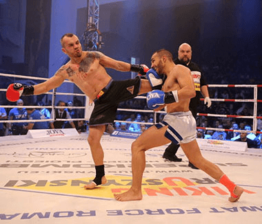

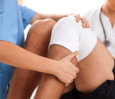
The knee is one of the most frequently affected joints in contact sports, such as soccer and rugby, but also in sports that require rapid changes of direction, such as basketball, football, and volleyball. Numerous specialized studies describe the functional importance of meniscal tissue, namely: shock absorption, stabilization, proprioception, lubrication, and nourishment of the knee joint.
The medial meniscus covers approximately 64% of the internal tibial plateau, while the lateral meniscus covers 84% of the external tibial plateau. In flexion, the menisci move forward and backward, and in extension, the reverse occurs. According to specialized studies, the medial meniscus has about 5 mm of movement, while the lateral meniscus has 11 mm of movement from anterior to posterior, during knee flexion between 0-120°. A smaller amount of movement occurs at the posterior horns of the menisci, which makes them more susceptible to shear forces if there is a ligament deficiency in the knee. The peripheral edge of the meniscus (10%-30% of the medial meniscus edge and 10%-25% of the lateral meniscus edge) is relatively well vascularized, which has important implications for its regeneration. This meniscal excursion, combined with reduced vascularization in the inner third of the meniscus, has led over the years to varied approaches in surgical technique and postoperative recovery.
It is widely accepted that meniscectomy induces degenerative changes in the knee. Following meniscectomy, short-term cartilage degeneration has been reported, especially in high-performance athletes. Therefore, the treatment strategy aims to preserve as much meniscal tissue as possible, and meniscus repairs and partial meniscectomies have replaced total meniscectomies. Meniscus repair techniques have advanced significantly, from open procedures to semi-open ones, and later to arthroscopic surgery.
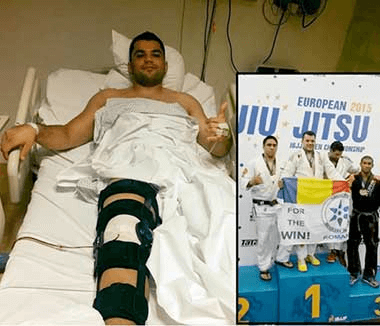
As with many other orthopedic procedures, the quality of postoperative recovery has a remarkable effect on the return to sports activity. An excellent surgical intervention followed by inadequate physical recovery will delay the resumption of activity or even create a potential risk of re-injury. An interdisciplinary team of specialists will contribute to the athlete's reintegration and to bringing them to a performance level similar to the preoperative one.
There are two different approaches to the postoperative recovery protocol: a protective model and an accelerated one. Protective protocols suggest that exercises be performed in an open kinetic chain (without weight bearing) and knee flexion below 90° in the first 6 weeks. Accelerated protocols allow early weight bearing and a range of motion with a variable progression of muscle strength.
The specialized literature describes a multitude of variables in decision-making during the recovery process, such as: the type of injury, its location, tissue quality, surgical technique, concomitant injury, and the involvement of both the patient and the therapist in the progression of the recovery protocol.
Longitudinal and "bucket-handle" tears located near the outer third (the vascular zone) or at the periphery of the meniscus have a faster recovery process compared to complex, horizontal, and radial tears, or those extending toward the center of the meniscus, in the middle third (the avascular zone).
It is imperative that there be a good working relationship between the surgeon and the therapist, to ensure communication of the surgical details, with the goal of individualizing each recovery protocol. Nevertheless, the principles of the recovery program should be clearly defined. Advancing from one phase of the recovery protocol to another is not necessarily chronological; this progression is based on meeting specific criteria, depending on the objectives of each recovery phase. Each phase should focus on the objectives set, and only once these objectives are met can the patient move on to the next phase of the recovery protocol.
During the first 6 weeks after surgery, the main goal is proper healing, which requires limiting stress on the joint and shear forces on the affected meniscus. At the beginning of this phase (day 1 - week 3), the recovery goals are pain control, edema reduction, neuromuscular re-education of the quadriceps, improving proprioception, and special attention is given to achieving complete passive knee extension. Hyperextension should be avoided in the case of an anterior horn suture. Patellar mobilizations should also be performed to prevent hypomobility.
The range of motion of the knee joint usually varies depending on the surgeon's preferences, taking into account intraoperative findings. As reference ranges, for peripheral meniscal injuries, the following are specified: 0-90° by the end of week 1, 0-100° in week 2, 0-120° in week 3, and 0-135° (passive) by week 4; these values are indicative, taking into account the patient's pain response and avoiding effusion. For complex posterior injuries, the range of motion is limited to 0-70° for 3 weeks, progressing to 0-90° in week 4 and to 120° in week 5. Weight bearing is another variable component of postoperative recovery protocols.
In the case of simple peripheral sutures, for which an accelerated recovery protocol will be used, weight bearing is achieved progressively, with the knee immobilized in extension, so that by week 6 the joint can be fully loaded, with normal walking. In the case of complex sutures, the knee joint will not be loaded for 6 weeks, this being the protective period of recovery. Factors such as body mass index, the location of meniscal injuries, tissue quality, patient adaptability, and possible existing conditions all play a role in decision-making for establishing the recovery process, from gradual joint loading to full loading.
It is important to mention that the semimembranosus muscle also inserts on the posterior horn of the medial meniscus, and the popliteus muscle on the lateral meniscus; therefore, care must be taken with hamstring exercises for 6 weeks postoperatively. An upper body ergometer can be used to maintain the physical condition of a patient who cannot load the knee joint or who has a mobility restriction. The cycle ergometer should not be added until the patient can easily achieve 110° flexion and should not be used to gain mobility because of the shear forces exerted on the knee.
To move on to the next stage of the rehabilitation process, the patient must have complete passive range of motion, no effusion, normal gait, and neuromuscular control of the lower extremity. Quadriceps control, as well as knee stability in the frontal and sagittal planes, should be good, and the patient must have adequate confidence to progress with a closed kinetic chain program.
The range of motion in complex meniscus tears may remain at 0-120° for the first 2 weeks of this phase (weeks 7 and 8 postoperatively), in order to further promote tissue healing and protect the joint, in situations where the tissue is friable or there are other intraoperative issues. Gait and balance re-education will be done at the start of this phase; proprioceptive training will be initiated starting with weight shifting. One can progress with closed kinetic chain exercises, with front or lateral step-ups and step-downs. Later, unilateral work can be done, within the range of motion in which the joint is protected. Emphasis should be placed on single-leg support balance - proprioceptive activities. Swimming can be used as a tool for conditioning. However, swimming with the help of a float, the crawl stroke, or backstroke should be the only ones practiced, since the butterfly stroke involves significant knee varus and valgus at certain points of the movement, which produces potential shear forces that could affect the repair process.
The first goal of the return-to-activity phase is to improve muscle strength up to the preoperative level. Neuromuscular training continues, and later progresses from light jumps to a running program with controlled intervals, also aiming to improve cardiovascular endurance. The goal of this phase is to ensure adequate explosive strength, elastic or reactive strength, and speed, with excellent knee mechanics. Activities are performed that mimic fatigue and the demands associated with the sport practiced, but in a controlled environment, in order to continue protecting the postoperative repair. Assuming the patient has progressed without complications, the difficulty level of exercises for increasing muscle strength can be raised at this stage (increased intensity with fewer repetitions).
Activities performed in a closed kinetic chain can have a higher degree of difficulty in ranges of motion below 90°. In complex tears, closed kinetic chain exercises such as squats or leg presses should remain above 90° until after 14 weeks. Care must be taken to avoid patellofemoral pain or symptoms of patellar tendon overload, and loads should be progressive.
Aerobic endurance continues to be a goal during the return-to-activity phase, for which the cycle ergometer and the stair master (stair-climbing machine) can be used. It is important to monitor compensations and pay attention to cumulative loads on the patellar tendon, and to avoid excessive patellofemoral loading associated with fatigue during recovery activities.
.jpg)
Many surgeons recommend running starting at weeks 12-16 postoperatively for peripheral tears, and at weeks 16-24 for complex tears. The gradual resumption of running, with the surgeon's permission, is done provided that the patient has good control in the frontal and sagittal planes, adequate muscle endurance, and can perform jumps and agility activities with good physical condition and no symptoms. The return to running must be carried out in a controlled setting, starting with interval running and a slow, controlled progression of distance (treadmill); this will vary depending on the type of sports activity practiced. In this phase, resistive lateral exercises can be added with careful monitoring, in order to avoid external tibial rotation.
After successfully resuming running, one can progress to a plyometric program, taking care, however, since the ground reaction forces experienced during such activities place very high stress on the anatomical structures. During jumps, the ground reaction force can be 4-11 times greater than body weight.
As has been extensively described in the specialized literature regarding return to play following anterior cruciate ligament (ACL) reconstruction, there is no single test, but rather a series of tests that have proven effective in helping decide on an athlete's return to activity. With regard to meniscus repair, in ideal cases (injury in the vascular zone, ideal tissue, excellent fixation, and an aggressive weight-bearing program), return to activity is granted at 4-5 months. Obviously, in complex injuries or in situations where other factors contribute to the healing issue, return to activity can take up to 6 months or longer.
Different methods have been used to interpret postoperative results. These have included patient history, clinical examination, diagnostic tests such as arthrography or magnetic resonance imaging (MRI), and evaluation through arthroscopic control. Arthrography or MRI have not been precise in assessing the postoperative state of the meniscus. These imaging studies have limited sensitivity in determining tear patterns, the actual size of the tear, the portion of the tear that has not healed, or degeneration in a meniscus after surgery. Arthroscopy is the most objective method for evaluating healing following meniscus repair.
The complete healing rate following the procedure is approximately 60%, according to the specialized literature. However, it has been shown that patients whose meniscus is incompletely healed can remain asymptomatic. According to a study of 140 patients who underwent simultaneous anterior cruciate ligament (ACL) reconstruction, complete healing of the meniscus repair was reported in only 84.3% at arthroscopic follow-up, while the clinical success rate was 96.4%. This is because some patients in the completely healed group did not present symptoms related to the meniscus.
The term "satisfactory meniscus healing" has been introduced in the specialized literature, covering both complete healing and an asymptomatic meniscus. Several studies have concluded that there is no decline in results over time, not even for a partially healed meniscus, reporting that it retains a biomechanical function that protects the articular cartilage from degenerative changes.
A meniscus repair is considered a clinical success when there are no residual or recurrent symptoms or mechanical pain related to the meniscus, and no subsequent surgical procedures are performed on the repaired meniscus. Good to excellent results have been reported in most long-term studies, with a success rate ranging between 59% and 100%. Following a systematic review of studies conducted over a period longer than 5 years, the overall clinical success rate was 76.9%.
The knee that has undergone surgery is usually protected during the early postoperative period to promote healing. Immobilization and disuse of the joint affect knee function, and the athlete must regain muscle control, proprioception, and confidence in using this joint.
Athletes represent the most demanding subgroup of patients, mainly for 2 reasons. First, they have high expectations of returning to their preoperative level of activity; second, they place the greatest load on the meniscus and, as a result, place maximum stress on any meniscal reconstruction. Thus, during preoperative planning it is useful for both the surgeon and the patient to have realistic expectations regarding the surgical success of the meniscal procedure and the likelihood of resuming that particular sports activity.
In general, it appears that most patients manage to resume their sports activity following meniscus repair. The results of recent studies regarding the return to play (RTP) rate show:
The time from surgery to resuming activity can play a significant role in decision-making when it comes to an athlete with a meniscus tear. In the specialized literature, the average time for a patient who has undergone a meniscal procedure to be able to fully participate in sports activities ranges between 4 and 6 months.
Another important variable is whether the patient will be able to return to their preoperative level of activity or to a lower level following meniscus repair. Meniscus repair is associated with a return to a high level of activity, which can be the same as or at least close to the preoperative level. An initially high rate of return to activity (RTP) following meniscus repair or the level of sports activity may decrease over time, in long-term follow-up. However, this decrease in RTP rates or activity level is more related to the patients' personal lives than to the meniscus recovery itself.
These are mainly related to the patient's characteristics, the timing of the injury, the nature of the meniscus tear or concomitant injuries, the patient's activity level, surgical techniques, and postoperative recovery.
The patient's gender and BMI have not been shown to be significant factors for the success of meniscus recovery.
It has been found that meniscal tissue contains more cells and has a better healing capacity in younger people. As a result, one might expect better results after meniscus repair in younger patients, but the specialized literature is not clear on this point.
A significantly higher success rate has been reported in younger patients. In a study that included only patients aged 17 or younger, the success rate was 100%. Another study, with a 10-year follow-up, compared meniscus repairs in patients younger than 40 with those aged 40 or older. They found no difference in the success rate or the overall outcome. Other authors reached the same conclusion, further noting that the higher demands in sports and profession in the younger population could compromise the outcome, despite a potentially better intrinsic healing capacity. In any case, in younger patients, preserving the meniscus should be the preferred option in order to reduce the risk of later osteoarthritis.
Age increases the likelihood of encountering a meniscal injury that is no longer suitable for repair, and factors such as osteoarthritis, which causes changes in joint alignment, must also be taken into account.
The time from injury to surgery appears to play a role in the healing and overall outcome of meniscus repairs. Many studies have shown that healing rates are improved when the procedure is performed closer to the time of injury, especially when it is done earlier than 6-8 weeks after the injury. Early intervention is encouraged, since chronicity can lead to deformation of the fragments, as well as secondary tears, which makes the fragments or the peripheral edge unsuitable for repair.
In athletes, early surgical intervention on a meniscus tear after injury not only has advantages for healing, but also avoids the post-traumatic consequences on the affected lower limb (muscle atrophy, loss of proprioception).
A meniscus injury has certain characteristics that the surgeon must take into account when deciding whether or not to perform surgery. These include the type of meniscal injury and the location of the tear in the medial or lateral meniscus, characteristics that may have prognostic value for meniscus healing following meniscus repair.
The types of injuries believed to be most likely to be repaired successfully are vertical longitudinal tears, bucket-handle tears, and meniscocapsular separations. In contrast, in the case of complex injuries, such as degenerative, radial, horizontal-plane tears, or tears with multiple "flaps," and oblique tears, which can extend from the vascular zone to the avascular zone, repair can be problematic. One study shows that the reoperation percentages for the symptomatic tibiofemoral joint were: 12% for single longitudinal tears, 27% for complex multi-plane tears, 27% for radial tears, and 29% for horizontal tears.
From a systematic review of studies on outcomes for repaired horizontal tears, it appears that their success rate is comparable to the success rate of other procedures for other types of meniscal injuries. According to some research, a success rate of 79% was found, at a follow-up over an average period of 40 months after meniscus repair for horizontal injuries; a significant decrease in functional outcomes was demonstrated in patients over 30 years of age. Typically, return to sports activity was not allowed earlier than 6 months postoperatively.
Partial radial injuries are usually problematic because they often affect the central avascular portions, which limits the chance of repair. In these cases, especially in small radial injuries, debridement of the floating edges is usually sufficient because the important circumferential fibers are largely intact and meniscus function is preserved.
The influence of tear length on the failure rate is a controversial subject. Certain authors have found no relationship between tear length and healing speed. In one study, tears of 3-4 cm showed a higher failure rate, but compared to smaller tears, the difference was not significant. Other authors have reported a substantially lower healing rate for tears larger than 4 cm (50%) compared to the healing rate for tears smaller than 2 cm (90%).
Regarding the location of the injury, it has been reported that injuries extending from posterior toward the center indicate a better healing rate compared to those isolated to the posterior area of the meniscus. In addition, although some studies could not observe any significant difference in outcomes after medial meniscus repair compared to results obtained after lateral meniscus repair, it appears that, in long-term studies, there is a trend toward a slightly lower failure rate for lateral meniscal procedures. Also, the potential sequelae of meniscectomy are more severe for the lateral meniscus, and therefore, in the decision-making process, it matters which of the menisci is affected.
Two separate studies reported a large decrease in healing time if the margin width exceeded 4 mm from the meniscosynovial junction. However, an extension of the injury into the avascular zone is not a contraindication for surgery. Procedures for injuries extending into the avascular zone have had good results, especially in young athletes, since there is not only an increased healing capacity, but the chance of healing likely outweighs the potential risks of the procedure.
Attempts have been made to promote bleeding in the avascular zone of the meniscus, with some surgeons using exogenous fibrin clots to enhance recovery and promote healing of injuries located in the avascular zone. This technique has been successful and has been associated with a return to preoperative performance levels or close to the previous level. Other clinical applications that enhance meniscal recovery and can promote healing include microfracture of the intercondylar eminence and platelet-rich plasma.
There is an ongoing debate regarding the positive effect of simultaneous ACL reconstruction on the healing of the operated meniscus. Many studies have reported higher healing rates following meniscus repair combined with ACL reconstruction, compared to an isolated meniscus repair without ACL injuries. The explanation is well known and is based on the hemarthrosis generated during ACL reconstruction, which provides the joint cavity with serum factors and fibrin clots that enhance the meniscus's healing response. On the other hand, several authors have found no positive effect of ACL reconstruction on the success rate of meniscus repair.
Patients with isolated meniscal procedures, as mentioned earlier, usually return to activity between 4 and 5 months postoperatively. However, simultaneous ACL reconstruction increases the period from surgery to return to activity. Most authors allow patients who have undergone meniscus repairs together with ACL reconstruction to participate in sports activities after 6 months postoperatively, if certain clinical goals have been met. Moreover, it has been found that the postoperative activity level for patients without ACL reconstruction is significantly higher (average Tegner score of 8.00) than that for patients with ACL reconstruction (average Tegner score of 6.8).
If the surgical technique can affect the healing response, then the long-term outcome may also be a concern for the surgeon. Regarding suture orientation, in general, vertical sutures are preferred over horizontal sutures, since these result in more rigid repairs. Today, open meniscal procedures are rarely performed. Indications may include a very tight medial compartment or concomitant pathologies that must be addressed through an incision, such as meniscal cysts. High healing and return-to-activity rates have been reported following open meniscal surgery for horizontal meniscal injuries in young patients, over an average period of 4 years. However, it appears that open surgery requires a more conservative rehabilitation protocol and delays the resumption of sports activity.
The most controversial factors in recovery protocols are immobilization and weight-bearing status. A recent systematic review on this topic concluded that early joint mobilization and immediate postoperative weight bearing do not influence clinical outcomes after isolated meniscectomy.
Current evidence suggests that recovery programs should be individualized, taking into account the "personality" of the injury, aiming for an accelerated recovery and return-to-activity process, while avoiding harmful effects on the healing of the repaired meniscus. Two different recovery protocols have been established: an accelerated program for peripheral meniscus injuries and a conservative program for complex injuries. In the long term, meniscus repair is associated with a high success rate in athletes returning to their activity, ranging between 59% and 100%. The postoperative return-to-sports-activity rate (RTP) is usually the same as or similar to the preoperative level. When isolated procedures are performed, return to activity occurs in approximately 5 months, whereas when combined with ACL reconstruction, 6 months or more are required. There is strong evidence to support meniscus repair over meniscectomy in decision-making regarding meniscocapsular separation, longitudinal tear types, bucket-handle tears, and those located in the outer two-thirds of the meniscus. Other factors that have been associated with a superior outcome following meniscus repair include younger patient age, a period of less than 6-8 weeks between trauma and surgery, a shorter length of the meniscus tear, location of the injury in the medial meniscus, and simultaneous anterior cruciate ligament reconstruction.
Centrokinetic is the place where you will find clear answers and solutions for your movement problems. The clinic dedicated to musculoskeletal conditions is divided into the following specialized departments:
Find out the latest news by following the Centrokinetic clinic's Facebook and YouTube accounts.
See details
READ MORESee details
READ MORESee details
READ MORESee here how you can make an appointment and the location of our clinics.








CENTROKINETIC FLOREASCA
Bucharest, 1st District,
Mircea Eliade Street, no. 18,
Eliade Tower, Entrance A, Floor 3
| Navigation | Explore 3D |
CENTROKINETIC Barbu Vacarescu
Bucharest, 2nd District,
Barbu Vacarescu Street, no. 102,
102 The Address, ground floor.
| Navigation | Explore 3D |
CENTROKINETIC MILITARI
Bucuresti, Sector 6,
Bld. Iuliu Maniu, nr. 15G,
Cladirea PBT, etaj 3.
| Navigation |
CENTROKINETIC CLUJ-NAPOCA
Cluj-Napoca,
Str. Intre Lacuri, nr.1
Intrarea este in spatele cladirii.
| Navigation |
SOCRATES CLINIC TIMISOARA
Timisoara, Calea Martirilor 1989,
nr. 1.
Telefon: 0371 785 374.
| Navigation |
Phone: 0319693
E-mail: programari@centrokinetic.ro
Operating hours:
Monday – Friday: 07:00 – 21:00
Saturday: 09:00 – 14:00
Sunday: closed.
MAKE AN APPOINTMENT