
MRI or X-Ray: Which Investigation Gives You the Information You Need?


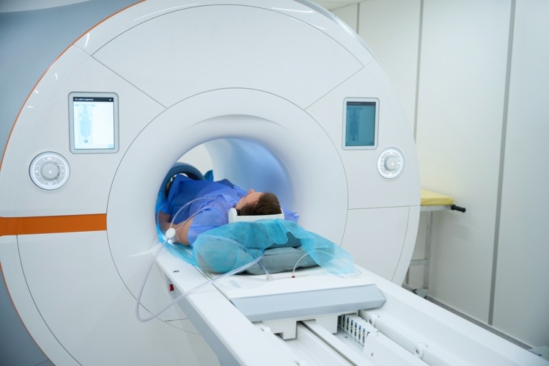
Does your knee hurt after a workout, did you have a fall, or did you receive a referral for imaging? In situations like these, it's natural to wonder which scan will give you the answer you need. Choosing between an MRI and an X-ray isn't random — it depends on your symptoms, the mechanism of injury, and the doctor's clinical suspicion.
A correct diagnosis starts with a clinical evaluation, and the right imaging test completes the information gathered during the consultation. Below, we explain when each option is useful and what questions are important to ask your doctor.
What is an X-ray and when is it useful?
An X-ray uses radiation to produce clear images of bone structures. Doctors recommend it frequently when there's suspicion of a fracture, dislocation, alignment changes, or arthritis. In orthopedics, an X-ray is often the first investigation ordered after an injury, or when pain appears suddenly and a bone problem needs to be ruled out.
Common reasons for an X-ray
Your doctor may order an X-ray if:
- you've had a fall and may have a fracture or dislocation;
- you feel intense pain after a sports injury;
- there's suspicion of scoliosis or other spinal deformities;
- the position of a prosthesis or implant needs to be checked after surgery.
Advantages of X-rays
- a quick investigation, usually taking 5-10 minutes;
- lower cost;
- wide availability;
- fast results in emergencies.
For recent injuries, an X-ray helps the doctor determine whether immobilization, surgery, or rest is needed.
Limitations of X-rays
X-rays don't adequately show:
- ligament tears (for example, an ACL tear);
- meniscus injuries;
- muscle tears;
- herniated discs;
- early inflammation or bone edema.
If pain persists and the X-ray shows no changes, the doctor may recommend a further investigation to assess the soft tissues.
What is an MRI and what information does it provide?
Magnetic resonance imaging uses a powerful magnetic field and radio waves to obtain detailed images across multiple planes. It doesn't use ionizing radiation. Through an MRI, the doctor can examine both bones and soft tissues: ligaments, tendons, muscles, intervertebral discs, or nerves.
When might a doctor order an MRI?
.jpg "e1343d318a175d4e8c297de237470f536e")
In most cases, the doctor recommends an MRI if:
- you have persistent joint pain with no clear cause on X-ray;
- you've suffered a ligament injury (ACL, PCL, collateral ligaments);
- there's suspicion of a meniscus tear;
- you have lower back pain radiating down the leg;
- you're recovering after orthopedic surgery and the doctor wants to assess the tissues.
Advantages of MRI
- doesn't use radiation;
- provides detailed images of soft tissues;
- identifies injuries at an early stage;
- allows precise treatment planning.
Read more about how magnetic resonance imaging works for more details.
Limitations and contraindications
An MRI takes longer, usually between 30 and 80 minutes. It's not recommended if you have:
- an incompatible pacemaker or cardiac defibrillator;
- certain ferromagnetic metal implants;
- metal fragments in the eye.
Always tell your doctor about any implant. Most modern titanium prostheses are compatible, but checking beforehand is still necessary.
MRI vs. X-ray - a direct comparison
To make the differences easier to understand, take a look at the table below:
| Criterion | X-ray | MRI |
|---|---|---|
| Energy type | X-rays | Magnetic field + radio waves |
| Radiation | Yes, low dose | No |
| What it shows best | Bones, fractures, alignment | Ligaments, tendons, discs, nerves |
| Duration | 5-10 minutes | 30-80 minutes |
| Cost | Lower | Higher |
| Typical indications | Acute injuries, arthritis | Ligament injuries, herniated discs |
| Contraindications | Limited | Incompatible metal implants |
The orthopedic doctor decides which option is appropriate based on the clinical exam and diagnostic suspicion.
Complementary investigations: when does ultrasound help?
In some situations, the doctor may recommend a musculoskeletal ultrasound. This investigation allows dynamic assessment of tendons and muscles. Ultrasound is useful for:
- superficial muscle injuries;
- tendinitis;
- monitoring the progress of a partial tear;
- guiding injections.
For subcutaneous masses or local inflammation, the doctor may recommend a soft tissue ultrasound.
Why does a correct evaluation and early intervention matter?
An early diagnosis gives you better chances of recovery and reduces the risk of complications. Many patients delay their consultation, hoping the pain will go away on its own. Sometimes it does, but often the condition progresses.
Benefits of a correct evaluation:
- the real cause of the pain is identified;
- unnecessary treatments can be avoided;
- proper recovery can start quickly;
- the risk of secondary injuries is reduced.
For patients in post-surgical recovery, imaging monitoring allows the physiotherapy and kinetotherapy program to be adjusted as needed.
Personalized approach and Centrokinetic's medical expertise
Every patient has a different history. A competitive athlete with a ligament tear has different needs than an adult with early-stage arthritis or a child with suspected scoliosis.
The Centrokinetic medical team carefully analyzes:
- symptoms;
- trauma or surgical history;
- activity level;
- mobility goals.
Based on this information, the specialists recommend the right investigation and build a personalized treatment and recovery plan.
Act early. Schedule a consultation at Centrokinetic, discuss it with the doctor, and follow the recommendations you receive.
Frequently asked questions
Can I get an MRI if I have a dental implant or braces?
In most cases, yes. Modern dental implants are compatible with MRI. Inform the doctor before the investigation to check the type of material.
Does repeated X-ray exposure affect your health?
The radiation dose required for a standard X-ray is small. The doctor only recommends the investigation if the benefit outweighs the risk. For frequent monitoring, the specialist may opt for radiation-free methods, if the situation allows it.
How long does an MRI take and how do I prepare for it?
An MRI takes between 30 and 80 minutes, depending on the area being examined. Usually, no special preparation is needed. You'll receive clear instructions before your appointment.
Can I have an MRI or X-ray during pregnancy?
X-rays are avoided, especially in the first trimester. An MRI can be performed in certain situations if the doctor considers that the benefits outweigh the risks. Discuss it with your obstetrician and the radiologist before scheduling.
Disclaimer: This material is for informational purposes only and does not replace specialist medical consultation.
Sources:
MAKE AN APPOINTMENT
FOR AN EXAMINATION
See here how you can make an appointment and the location of our clinics.
MAKE AN APPOINTMENT
BUCHAREST TEAM


































































CLUJ NAPOCA TEAM


































BRASOV TEAM



















Asiguratori privati





Parteneriate



CENTROKINETIC FLOREASCA
Bucharest, 1st District,
Mircea Eliade Street, no. 18,
Eliade Tower, Entrance A, Floor 3

| Navigation | Explore 3D |
CENTROKINETIC Barbu Vacarescu
Bucharest, 2nd District,
Barbu Vacarescu Street, no. 102,
102 The Address, ground floor.

| Navigation | Explore 3D |
CENTROKINETIC MILITARI
Bucuresti, Sector 6,
Bld. Iuliu Maniu, nr. 15G,
Cladirea PBT, etaj 3.

| Navigation |



CENTROKINETIC CLUJ-NAPOCA
Cluj-Napoca,
Str. Intre Lacuri, nr.1
Intrarea este in spatele cladirii.

| Navigation |


SOCRATES CLINIC TIMISOARA
Timisoara, Calea Martirilor 1989,
nr. 1.
Telefon: 0371 785 374.

| Navigation |
CONTACT
Phone: 0319693
E-mail: programari@centrokinetic.ro
Operating hours:
Monday – Friday: 07:00 – 21:00
Saturday: 09:00 – 14:00
Sunday: closed.
MAKE AN APPOINTMENT

