
Open MRI vs. Closed MRI: Differences, Advantages, and Who It Is Recommended For in 2026


Open MRI vs. Closed MRI: Differences, Advantages, and Who It Is Recommended For in 2026
Have you been recommended an MRI and don't know whether to choose the open or the closed version? This is a common uncertainty, especially if you are dealing with back pain, a sports injury, or are recovering after an orthopedic procedure.
In 2026, imaging technology allows increasingly precise investigations. However, the right choice does not automatically mean the most powerful machine, but the solution suited to your medical problem. Here is what you need to know!
What is a closed MRI and how does it work?
The classic MRI, also called closed, uses a powerful magnet, usually 1.5 Tesla or 3 Tesla. The procedure requires you to enter completely into a cylindrical tube, so that the machine can capture detailed images of organs, joints, ligaments, and nerves.
The 1.5T or 3T MRI offers high resolution, useful in evaluating fine spinal cord lesions, brain pathology, or small tumors. Main advantages:
- Very good resolution for small structures;
- Shorter examination time (15–45 minutes, in most cases);
- Detailed imaging for neurological and oncological conditions;
- Standard frequently used in complex preoperative evaluations.
Limitations:
- Narrow space, which can trigger anxiety;
- Intense noise during the scan;
- Restrictions for people with a large body circumference.
For an athlete with a suspected complex ligament injury of the knee, the closed MRI can provide additional details useful to the surgeon. For evaluating a brain tumor or a fine spinal cord lesion, doctors almost always recommend this version.
What does an open MRI mean and how does it differ?
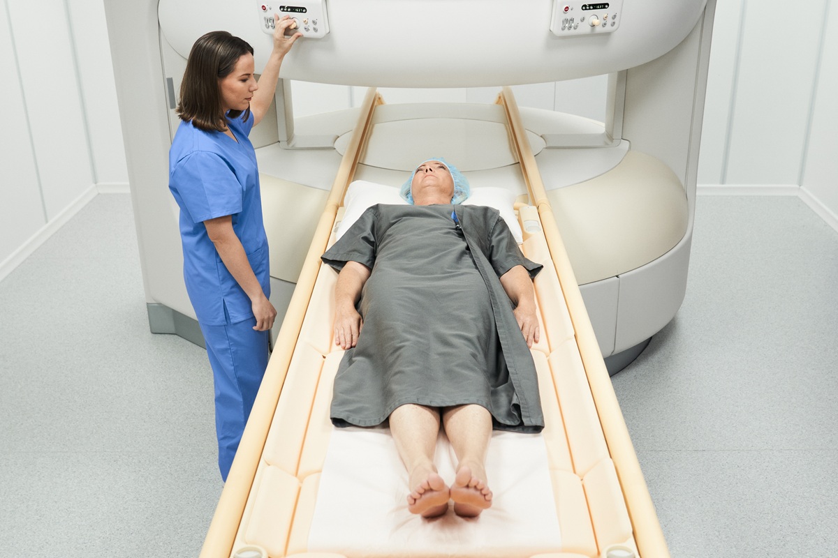
The open MRI has a different configuration. The magnets are positioned above and below or laterally, and the sides remain open. You do not have to enter a narrow tunnel; instead, you stay in an airy space, with visibility toward the outside.
The magnetic field is usually lower (0.2–1 Tesla). For this reason, the scan can take longer, between 30 and 60 minutes.
Common advantages:
- Increased comfort for people with claustrophobia;
- Easier access for overweight patients;
- More flexible positioning in case of intense pain;
- Lower noise.
Limitations:
- Lower resolution for very fine lesions;
- Longer examination time.
At Centrokinetic, MRI investigations are adapted to musculoskeletal pathology. At the dedicated location in Cluj, you can opt for an open MRI, frequently indicated for the lumbar spine, knee, shoulder, or ankle.
Direct comparison: relevant criteria for the right choice
| Selection criterion | Closed MRI | Open MRI |
|---|---|---|
| Magnetic field strength | 1.5T – 3T (high) | 0.2T – 1T (low) |
| Image quality | Very detailed; ideal for fine lesions | Good; optimized for orthopedic pathology |
| Examination duration | 15 – 45 minutes (faster) | 30 – 60 minutes (slower) |
| Psychological comfort | Can cause anxiety or claustrophobia | Well tolerated; a feeling of freedom and space |
| Common indications | Brain, spinal cord, oncology | Joints, spine, sports injuries |
| Accessibility | The standard in most clinics | Recommended for children or people diagnosed with obesity |
Who is the open MRI recommended for?
The open MRI, such as, for example, the Aperto Lucent MRI, is a suitable choice in several situations:
- You have claustrophobia and were unable to complete a closed MRI;
- You have intense low back pain and find it hard to stay still in a narrow space;
- You are an amateur athlete with a suspected meniscus tear or ligament injury;
- You need monitoring during orthopedic post-operative recovery;
- You are looking for an evaluation for common conditions such as back pain.
For parents seeking investigations for active children or teenagers, the open environment reduces stress and lowers the need for sedation.
When is the closed MRI preferred?
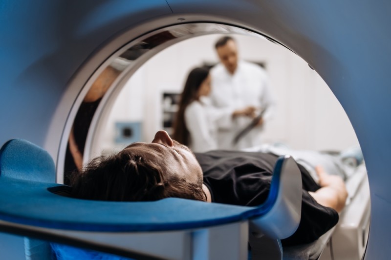
There are situations in which the doctor directly recommends the classic version:
- Suspected multiple sclerosis or demyelinating lesions;
- Evaluation of brain tumors or metastases;
- Subtle spinal cord lesions;
- Complex surgical planning;
- Spinal stenosis with multiple nerve compression.
In the case of a herniated disc with significant neurological deficit, the doctor may request high-resolution images to decide whether intervention is needed. Do not choose the type of machine without a specialist's recommendation. The radiologist and the orthopedic doctor together determine the appropriate investigation.
Safety, contraindications, and preparation
MRI does not use ionizing radiation. The investigation is considered safe for most patients. However, there are situations in which caution is necessary:
- Incompatible pacemaker;
- Old metallic implants;
- Intraocular metallic fragments;
- Severe kidney failure (if contrast agent is used).
Inform the medical team about any implanted device. Remove all metallic objects. If you feel nervous, discuss comfort options in advance.
A correct evaluation before the investigation prevents postponements and repeat examinations. Early intervention, based on clear images, helps establish a personalized treatment plan and can shorten the recovery period.
Why does the integrated evaluation at Centrokinetic matter?
For active adults and athletes, a diagnosis is not just the confirmation of an injury. What you do afterwards matters. In a center specialized in medical imaging, the radiologist correlates the images with the clinical examination. The orthopedic doctor or the recovery specialist establishes an adapted plan: physical therapy, injections, monitoring, or surgery, if necessary.
The personalized approach reduces the risk of unnecessary treatments and supports a safe return to daily or sporting activities.
Frequently asked questions
Is the open MRI sufficient for a herniated disc?
In most cases of lumbar herniation without severe neurological involvement, the open MRI provides adequate information for establishing the treatment. If symptoms such as muscle weakness or sensory disturbances appear, the doctor may recommend the classic version for more detailed images.
How long does the investigation take and when do I receive the result?
The closed MRI usually takes between 15 and 45 minutes. The open version can take up to 60 minutes. The written result is usually available within 24–48 hours, depending on the complexity of the case.
Can I have an MRI if I have a dental implant or a prosthesis?
Most modern dental implants and prostheses are MRI-compatible. However, inform the medical team and bring the device's documents. The doctor checks the type of material and decides whether the investigation can be performed safely.
If you have persistent symptoms, do not postpone the investigation. A timely diagnosis gives you more therapeutic options and increases the chances of an effective recovery. Book an appointment at Centrokinetic now for a specialist consultation!
Sources:
- Behluli, E., et al. "Patient-Centric Comparative Analysis of Experiences in Open Upright and Conventional Closed MRI Scanners." Radiography, vol. 30, no. 5, Aug. 2024, pp. 1258–1264. Accessed 7 Apr. 2026.
- Clinic, Cleveland. "An MRI (Magnetic Resonance Imaging) Is a Test That Creates Clear Images of Structures inside Your Body Using a Large Magnet, Radio Waves and a Computer." Cleveland Clinic, 8 Sept. 2023. Accessed 7 Apr. 2026.
MAKE AN APPOINTMENT
FOR AN EXAMINATION
See here how you can make an appointment and the location of our clinics.
MAKE AN APPOINTMENT
BUCHAREST TEAM


































































CLUJ NAPOCA TEAM


































BRASOV TEAM



















Asiguratori privati





Parteneriate



CENTROKINETIC FLOREASCA
Bucharest, 1st District,
Mircea Eliade Street, no. 18,
Eliade Tower, Entrance A, Floor 3

| Navigation | Explore 3D |
CENTROKINETIC Barbu Vacarescu
Bucharest, 2nd District,
Barbu Vacarescu Street, no. 102,
102 The Address, ground floor.

| Navigation | Explore 3D |
CENTROKINETIC MILITARI
Bucuresti, Sector 6,
Bld. Iuliu Maniu, nr. 15G,
Cladirea PBT, etaj 3.

| Navigation |



CENTROKINETIC CLUJ-NAPOCA
Cluj-Napoca,
Str. Intre Lacuri, nr.1
Intrarea este in spatele cladirii.

| Navigation |


SOCRATES CLINIC TIMISOARA
Timisoara, Calea Martirilor 1989,
nr. 1.
Telefon: 0371 785 374.

| Navigation |
CONTACT
Phone: 0319693
E-mail: programari@centrokinetic.ro
Operating hours:
Monday – Friday: 07:00 – 21:00
Saturday: 09:00 – 14:00
Sunday: closed.
MAKE AN APPOINTMENT

