See details
READ MORE

Osteoarthritis (OA) is a progressive degenerative disease that affects the articular cartilage, subchondral bone, synovium, and joint capsule. It has a multifactorial etiology and affects approximately 60% of people over the age of 50. According to Davis et al., OA affects 9% of men and 18% of women over 65 and is responsible for high levels of absenteeism and retirement due to disability.
The etiology of OA is related to repetitive mechanical loading and age. Recent studies have separated the etiological factors into three main subgroups: sex, anatomy, and body mass. The reduction in strength in the muscle groups involving the joints is significant, as it causes progressive loss of function.
These symptoms significantly restrict the individual's ability to get up from a chair, walk, or climb stairs. During movements, crepitus may be heard due to arthritis of the irregular joint surfaces. OA often affects one of the three compartments of the knee: the patellofemoral joint, the medial compartment, or the lateral compartment. In relation to severity, knee OA can be classified according to the Kellgren-Lawrence scale, which consists of five grades: 0 = no OA, 1 = doubtful, 2 = minimal, 3 = moderate, and 4 = severe.
Treatment of OA should be multidisciplinary and aim at functional and clinical improvement.
Physiotherapy is a non-pharmacological intervention for knee osteoarthritis, recommended by the American College of Rheumatology and the European League Against Rheumatism. A systematic review has shown that physical exercise reduces pain and improves functionality in patients with knee OA. One of the most common ways of achieving this functional improvement is following a daily exercise program. Guidance from the physical therapist ensures that the exercises are performed correctly. Unfortunately, there is high demand on large public health services, and it is often impossible to meet everyone's needs, especially those who have difficulty traveling to the recovery center. However, physical exercises can be carried out both at the clinic and in home exercise programs, provided that the patient returns for certain supervised exercises.
The usefulness of home exercises was clearly shown by another study conducted by Deyle's research team, which compared a group of OA patients undergoing exercises, individualized manual therapy, and a home exercise program for four weeks with a group of OA patients receiving the same home exercise program and a clinic visit 2 weeks later. Both groups showed significant clinical improvement compared to baseline. Initially, the patients at the clinic showed a higher level of improvement, but the results of both groups were equally beneficial one year later. In addition, another study showed that home exercise for two years could lead to significant control of knee pain. Thus, the objective of this study was to evaluate the effectiveness of a physical activity guidance manual to be performed at home by patients with knee osteoarthritis. The study evaluated pain, joint range of motion, muscle strength, and functional capacity before and after treatment. The hypothesis was that OA patients would benefit from physical therapy at home, with the help of the manual, and would show improvements in pain, strength, joint range of motion, and functional capacity.
This observational study was carried out in the second half of 2007 at the rehabilitation service of a public university hospital (Santa Casa de Misericordia de Sao Paulo). This was a prospective case series on the application of a physiotherapy intervention (guidance manual) for patients with knee osteoarthritis (OA). Pain, joint range of motion, strength, and functionality were assessed before and after the application of the physiotherapy intervention.
This study included all patients diagnosed with grade 2 or 3, according to the Kellgren-Lawrence scale, and patients with mild to moderate osteoarthritis, who were referred to physical therapy at a public university hospital. Both men and women (45 years old) were recruited for this study. Patients had to have been referred by physicians (medical specialists), and all underwent X-rays. Patients with grade 1 or 4 on the Kellgren-Lawrence scale were excluded. In addition, we also excluded patients who had undergone any type of orthopedic surgical intervention on the lower limb in the last two years and/or other concomitant therapies, except for the use of analgesic and non-steroidal anti-inflammatory drugs. During the study period, 98 patients were included in the study.
All patients who came to the physiotherapy service came for an initial evaluation visit. During the first visit, the following data were collected: quantitative assessment of pain using the visual analog scale, joint range of motion and muscle strength, qualitative assessment of the main affected functions, and the duration for which they had experienced pain (in months or years).
After the clinical evaluation, patients were divided into groups of up to five people. They were given an explanation of the disease and a program of stretching and muscle-strengthening exercises, to be performed individually, at home. Patients performed each exercise in the presence of the physical therapist, so that any questions about the exercises could be clarified. All patients received a printed manual written in simple language that contained photographs and instructions. Patients were reassessed at monthly intervals for at least 90 days from the initial assessment. In these reassessment sessions, the physical therapists observed how the patient performed the exercises at home. If the patient had no difficulty performing the exercise, then the number of repetitions was increased to a maximum of five repetitions in each set. All patients participated in the follow-up sessions.
To assess the intensity of knee pain, the visual analog scale was used: zero (0) indicated the absence of pain and 10 indicated unbearable pain intensity. Pain was assessed before treatment and at the end of it. Manual muscle strength tests were applied to the extensor and flexor muscle groups of the knee, using the Kendall scale, and grades ranged from zero (no contraction) to five (maximum strength). The same evaluator performed all assessments before and after the protocol. The Lequesne algofunctional questionnaire for knee and hip osteoarthritis was applied at the initial evaluation and at 90 days. The questionnaire results in the construction of the Lequesne algofunctional index. The patient answered questions regarding the knee joint with the following scores: extremely severe (equal to or greater than 14 points), very severe (11 to 13 points), severe (8 to 10 points), moderate (5 to 7 points), or mildly affected (1 to 4 points).
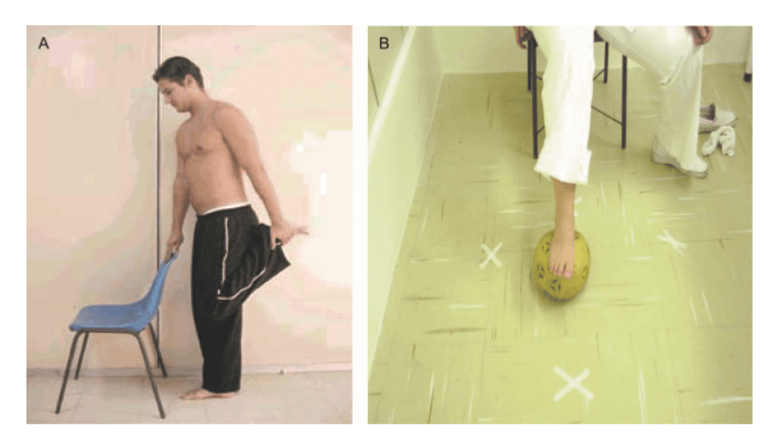
Figure 1 - Examples of two exercises proposed in the manual and their accompanying photographs.
(A) While standing, hold onto a chair or table to maintain your balance, bend one of your knees and with your hand, pull your foot toward your buttocks. If you cannot reach your foot, pull it with the help of a folded towel.
(B) Mark the floor with adhesive tape in different directions. Sit on a chair, step on a ball, and roll it to the marks made on the floor. Repeat 10 times.
At the beginning, it gave a definition of OA and general anatomical aspects of the knee, including diagrams. It then emphasized the importance of daily exercises for improving symptoms and functionality. This first part of the brochure also contained some important tips for everyday life (for example, not putting too much strain on the knee joint by distributing heavy household tasks throughout the week, maintaining an ideal body weight, avoiding wearing high heels, and avoiding activities that involve high impact on the joint). Some simple analgesic measures that could easily be applied at home were also addressed, including cryotherapy and compression through local application for a maximum of 20 minutes. Next, the brochure presented stretching exercises (hamstrings, gluteal muscles, quadriceps, and triceps surae) and muscle-strengthening exercises (quadriceps, hamstrings, gluteus medius and maximus, adductor, and triceps surae) starting with some isometric exercises for the knee and isotonic exercises for the hips. Each of the 20 exercises was explained, and each exercise was accompanied by a photograph of a model performing the proposed movement. Figure 1 provides an example of the proposed exercises.
This study analyzed the visual analog scale (0 to 10), knee range of motion (0 to 140˚), the strength of the extensor and flexor muscle groups (0 to 5 points), and the Lequesne index (1 to 14 points or more). After data analysis, each of the variables was subjected to a standardization test to determine the types of statistical tests to be used. For quantitative variables, this analysis was done by observing the minimum and maximum values and by calculating the means and standard deviations. For patients with bilateral pain, the average value between the measurements of both knees was calculated. For patients who had OA in only one knee, the unaffected knee was not taken into account. The paired Wilcoxon test was used for VAS and ROM, which compared the data before and after the physiotherapy intervention. The paired t-test was used to analyze the results of the Lequesne Algofunctional scale. For qualitative variables, absolute and relative frequencies were calculated. The Kruskal-Wallis test was used to compare the differences between pre- and post-treatment according to the patient's age. For all analyses, differences were considered significant when p < 0.05. All analyses were performed using the Statistical Package for the Social Sciences (SPSS), version 15.0.
As already mentioned, during the study period, 98 patients with knee osteoarthritis were seen by the physiotherapy services (with a total number of 166 affected knees). All patients participated in the follow-up sessions, but it was not possible to apply the Lequesne assessment to all patients due to a lack of specialized staff. Therefore, the results presented here refer to a sample of 38 patients who were clinically evaluated and answered the Lequesne questionnaire. As shown in Table 1, 84% of patients were women, 50% were between 50 and 59 years old, and 66% had bilateral pain. During the initial evaluation, all patients reported knee joint pain during daily activities as the main complaint (for example, difficulty going up and down stairs, squatting, and kneeling). Table 2 shows that the exercise program led to significant changes in all measurements (VAS, ROM, muscle strength, and Lequesne assessments). All variables had average variations greater than 50% compared to baseline (p < 0.05). Patients were subdivided into three age groups for comparison. There was no association between pain and age, before or after treatment, and age was not associated with range of motion, muscle strength, or functional status after treatment either. Table 3 shows that age did not affect the results (p > 0.05); all patients benefited from treatment regardless of age.
Characteristics | Frequency | % |
Sex | ||
Female | 32 | 84.2 |
Male | 6 | 15.8 |
Age | ||
50-59 | 19 | 50 |
60-69 | 11 | 28.9 |
70+ | 8 | 21.1 |
Main problem | ||
Right knee | 8 | 21.1 |
Left knee | 5 | 13.2 |
Bilateral | 25 | 65.8 |
Total | 38 | 100 |
Variable | Deviation | SD | Median | Minimum | Maximum | N | p |
VAS pre | 7.87 | 1.12 | 8 | 5 | 10 | 38 | <0.01 |
VAS post | 3.58 | 0.92 | 3.5 | 2 | 5 | 38 | <0.01 |
ROM pre | 87.25 | 8.85 | 89 | 63 | 106 | 38 | <0.01 |
ROM POST | 123.71 | 7.6 | 124 | 107 | 137 | 38 | <0.01 |
MS hip Pre | 3.32 | 0.46 | 3.5 | 2 | 4 | 38 | <0.01 |
MS hip post | 4.78 | 0.38 | 5 | 4 | 5 | 38 | <0.01 |
MS Tendon pre | 3.7 | 0.39 | 4 | 3 | 4 | 38 | <0.01 |
MS Tendon post | 4.97 | 0.11 | 5 | 4.5 | 5 | 38 | <0.01 |
Lequesne Pre | 16.17 | 1.52 | 16 | 13.5 | 19.5 | 38 | <0.01 |
Lequesne Post | 7.57 | 2.19 | 7.75 | 4 | 12 | 38 | <0.01 |
Both immobilization and intense physical exercise can contribute to joint degeneration and increase the risk of developing the most severe form of OA, which requires arthroplasty, osteotomy, or arthroscopy. In a study conducted in the United States, the authors observed that strength, muscle contraction speed, and endurance were 30 to 50% lower in individuals with osteoarthritis compared to age-matched people without OA. OA leads to difficulties in carrying out daily activities and to a lack of physical activity as a result of pain. Weakness of the quadriceps muscle is considered one of the most important risk factors in the progression of knee OA. The strength of this muscle naturally decreases with age, which leads to functional limitation and possible degeneration of the knee joint cartilage. In our study, strength was assessed using the Kendall scale, which is a subjective measurement that is applied when dynamometers are not available. Although a significant increase in strength (25%) was observed after the home exercises, a greater change could probably have been detected with the help of dynamometry.
The most suitable non-surgical treatment for knee osteoarthritis (OA) is exercise therapy. Physiotherapy reduces pain and improves function, muscle strength, range of motion (ROM), joint stability, and aerobic conditioning. Regular practice of exercises performed by patients with knee osteoarthritis, in a program of intervention with appropriate guidance, can help, preventing the loss of muscle strength and the restriction of daily activities. In addition, regular exercise can allow pain control and can prevent loss of range of motion. Instructions on how to sit, lift, carry heavy objects, and walk correctly are useful and important in the treatment of OA. Strengthening exercises designed for the knee flexor musculature are just as important as those for the quadriceps in the rehabilitation of knee OA, due to the dynamic stability of this joint. Various studies have already shown good results with treatment protocols consisting of stretching the quadriceps, triceps surae, and dynamic resistance exercises of the hip and knee, with isometric and isotonic contractions. These exercises result in an improvement in joint range of motion, hip and hamstring strength, and dynamic activities, even when the exercises are performed in the patient's home. Compared to the suspension of physiotherapy after the patient's discharge, both isokinetic and isotonic exercises have shown good results in maintaining pain treatment at home following physiotherapy at a medical service center (assessed by the VAS score).
Functional capacity, assessed by the Lequesne Questionnaire, showed improvement in patients performing isokinetic exercises. In the guidance manual provided to the patients in the present study, the proposed strength exercises started with isometric training and then progressed to isotonic exercises. A literature search suggested that progressive isometric and isotonic exercises promote muscle strengthening and improved functionality. Studies have reported that the strength gain, which occurs through intramuscular adaptation, is achieved after 12 weeks of resistance exercise training. These findings justified the 90-day follow-up period in the present study, and we obtained excellent results. Increasing the amount of exercise stimulation (increasing repetitions and weight) is possible when the patient shows improvement or stability in OA signs and symptoms (that is, when pain, inflammation, and edema do not worsen or when these signs are progressively diminished). In this study, monthly visits allowed physicians to check the clinical status of each patient and to progress the exercises in order to determine the cases in which OA signs and symptoms had improved. In the present study, a significant reduction in pain and functional disability was observed according to the VAS score and the Lequesne Algofunctional Questionnaire, respectively. These results were similar to a study that found that a group of patients who performed isometric quadriceps exercises every day for eight weeks achieved a significant reduction in pain (assessed by VAS) compared to the control group, which was medicated with non-steroidal anti-inflammatory drugs. The improvement in function, assessed by the Lequesne Questionnaire, was also statistically significant (p = 0.001) in our study.
In the present study, pain was cited as the main complaint of all patients. Pain assessment is individual and subjective, since the individual is the authority on their own pain experience. The assessment was performed using the visual analog scale to identify pain intensity. In the reassessment that followed the procedure and physiotherapy guidance for performing the exercises, a statistically significant decrease in pain intensity was observed, which showed that the intervention had an effect on reducing pain. As an analgesic resource, local cryotherapy was always used in combination with compression and elevated positioning of the limb for approximately 20 minutes after exercises or when there was pain in the knee. Cryotherapy is a resource that is easy to apply at home. It is indicated in cases of pain, inflammation, and muscle spasms, since it acts directly on the nerve endings, which decreases impulse conductivity, causes an increase in the excitation threshold of nerve cells, favors a decrease in local cell metabolism, favors lower oxygen consumption, and prevents damage from secondary hypoxia. In order to maintain or improve the range of motion, stretching exercises were included in the manual given to patients. Joint mobility must be maintained or improved in patients with osteoarthritis, since loss of range of motion causes shortening and contraction of the muscles and capsular structures and can affect functionality. The individuals included in this study showed a statistically significant improvement in knee flexion range as a result of muscle stretching exercises. The short-term benefits of exercises for patients with knee OA levels 2 and 3 have been proven, such as pain reduction and functional improvement, but studies with a longer-term evaluation are needed, for example, nine months, as proposed by van Baar et al. In fact, it has been shown that short-term improvements can be seen with exercise programs for OA, but maintaining the benefits over a year depended on the patient's adherence to the protocol. Theoretically, maintaining the benefits can only be verified when periodic checks are performed, as proposed in the present study. When patients can be prescribed a home exercise program, more staff are available for other patients, especially those in acute conditions or surgical treatment. Thus, selecting patients who can benefit from a home exercise program is a cost-reduction measure; we believe that the results of this study can be obtained in any public service with similar patient characteristics: male or female, middle-aged or older patients with knee OA, who can read, and patients who do not have easy access to health services (a few reassessment sessions were needed to obtain positive results). The results would probably be even better in patients who have easier access to physical therapy supervision. However, our patients showed improvements in subjective symptoms, such as absence of pain, as well as objectively measured clinical status variables, such as function, flexion range of motion, and muscle strength, both for extensors and flexors.
Table 3 - Changes in pain measurements (analyzed by the visual analog scale, VAS), joint range of motion (ROM), muscle strength (MS), and functional status (by the Lequesne Algofunctional Questionnaire for knee osteoarthritis) according to age, and results of comparisons between age categories.
Variable | Age | Deviation | SD | Median | Minimum | Maximum | N | p |
VAS | 50-59 | 4.04 | 1.54 | 4 | 2 | 8 | 19 | 0.385 |
| 60-69 | 4.45 | 1.04 | 4 | 3 | 6 | 11 |
|
| 70+ | 4.63 | 1.06 | 4.5 | 3 | 6 | 8 |
|
| Total | 4.29 | 1.31 | 4 | 2 | 8 | 38 |
|
ROM | 51-59 | 38.95 | 8.67 | 40 | 30 | 53 | 19 | 0.097 |
| 61-69 | 35.5 | 7.76 | 34 | 36 | 51.5 | 11 |
|
| 71+ | 31.88 | 4.45 | 33.5 | 25 | 37 | 8 |
|
| Total | 36.46 | 8.04 | 35 | 20 | 53 | 38 |
|
MS hip | 52-59 | 1.55 | 0.47 | 1.5 | 1 | 2.5 | 19 | 0.514 |
| 62-69 | 1.41 | 0.63 | 1.5 | 0 | 2 | 11 |
|
| 72+ | 1.31 | 0.46 | 1.5 | 0.5 | 2 | 8 |
|
| Total | 1.46 | 0.51 | 1.5 | 0 | 2.5 | 38 |
|
MS tendon | 53-59 | 1.29 | 0.38 | 1 | 1 | 2 | 19 | 0.836 |
| 63-69 | 1.23 | 0.47 | 1 | 0.5 | 2 | 11 |
|
| 73+ | 1.31 | 0.37 | 1.25 | 1 | 2 | 8 |
|
| Total | 1.28 | 0.40 | 1 | 0.5 | 2 | 38 |
|
Lequesne | 54-59 | 8.32 | 1.25 | 8 | 7 | 11 | 19 | 0.514 |
| 64-69 | 8.95 | 1.84 | 8.5 | 6.5 | 12.5 | 11 |
|
| 74+ | 8.81 | 1.41 | 9 | 6.5 | 10.5 | 8 |
|
| Total | 8.61 | 1.46 | 8.5 | 6.5 | 12.5 | 38 |
The use of a home exercise guidance manual, together with monthly reassessment by a physical therapist, allowed patients with knee osteoarthritis to achieve significant improvement in pain, knee flexion, muscle strength, and functional capacity.
Centrokinetic is the place where you will find answers and clear solutions for your movement problems. The clinic dedicated to osteoarticular conditions is divided into the following specialized departments:
Find out the latest news by following the Facebook and YouTube accounts of Centrokinetic clinic.
See details
READ MORESee details
READ MORESee details
READ MORESee here how you can make an appointment and the location of our clinics.








CENTROKINETIC FLOREASCA
Bucharest, 1st District,
Mircea Eliade Street, no. 18,
Eliade Tower, Entrance A, Floor 3
| Navigation | Explore 3D |
CENTROKINETIC Barbu Vacarescu
Bucharest, 2nd District,
Barbu Vacarescu Street, no. 102,
102 The Address, ground floor.
| Navigation | Explore 3D |
CENTROKINETIC MILITARI
Bucuresti, Sector 6,
Bld. Iuliu Maniu, nr. 15G,
Cladirea PBT, etaj 3.
| Navigation |
CENTROKINETIC CLUJ-NAPOCA
Cluj-Napoca,
Str. Intre Lacuri, nr.1
Intrarea este in spatele cladirii.
| Navigation |
SOCRATES CLINIC TIMISOARA
Timisoara, Calea Martirilor 1989,
nr. 1.
Telefon: 0371 785 374.
| Navigation |
Phone: 0319693
E-mail: programari@centrokinetic.ro
Operating hours:
Monday – Friday: 07:00 – 21:00
Saturday: 09:00 – 14:00
Sunday: closed.
MAKE AN APPOINTMENT