See details
READ MORE

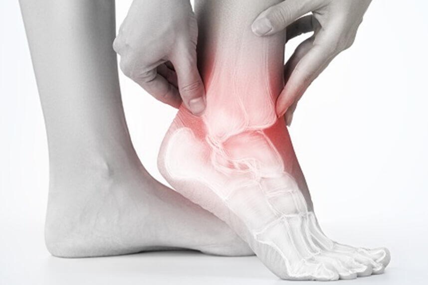
Centrokinetic meets patients' needs with a complete orthopedics department, a medical branch that deals with the diagnosis, treatment, and recovery of patients with conditions of the musculoskeletal system. These conditions are usually painful, acute or, most often, chronic, hindering the proper functioning of the musculoskeletal system.
Most of the time, the patient's evaluation is complex, multidisciplinary, given the systemic, generalized nature of most rheumatological conditions, which is why Centrokinetic has experienced doctors:
The sinus tarsi is a canal or tunnel formed between the talus and the calcaneus. Sinus tarsi syndrome is characterized by pain and inflammation in this area. Traumatic injuries to the ankle/foot (such as an ankle sprain) or prolonged exertion (such as standing or repetitive walking) are the main causes of this syndrome. It can also occur if the person has a flat foot or an overly arched, over-pronated foot, which can cause compression in the sinus tarsi area. Some characteristics are: pain in the anterolateral part of the ankle and a feeling of instability or difficulty walking on unstable surfaces.
Recent discussions about sinus tarsi syndrome (STS) describe this condition as primarily an instability of the subtalar joint due to ligamentous injuries, which results in synovitis and infiltration of fibrotic tissue into the sinus tarsi space.
The sinus tarsi is a tunnel between the talus and the calcaneus, which contains structures that contribute to ankle stability and its proprioception, but which can become damaged.
The joint between the talus and the calcaneus is also known as the subtalar joint or the talocalcaneal joint. This joint connects the lower surface of the talus and the upper surface of the calcaneus through their posterior articular facets, namely through the posterior calcaneal articular facet of the talus and the posterior talar articular facet of the calcaneus. Variation in the structure of these facets affects the stability of the subtalar joint.
This joint is also made up of the extrinsic ligaments (the calcaneofibular and deltoid ligament) and the intrinsic ligaments (interosseous, the talocalcaneal ligament, the cervical ligament, and the medial, lateral, and intermediate roots of the inferior extensor retinaculum. All the structures provide additional stability to the joint. Ruptures of the intrinsic ligaments allow increased mobility of the subtalar joint, which can lead to instability.
Anterolateral structures of the ankle:
Akiyama suggested that the sinus tarsi is not just a talocalcaneal joint space, but a source of nociceptive and proprioceptive information about foot and ankle movement, and that sinus tarsi syndrome may result from nociception and proprioception disorders in the foot.
Sinus tarsi syndrome mainly occurs after a traumatic ankle sprain or multiple ankle sprains (mainly due to a weak anterior talofibular ligament), which leads to injuries of the interosseous and cervical ligaments.
The ligaments can be partially or completely torn, and inflammation and hemorrhage of the synovial recesses in the tarsal sinus can occur. This happens in 70% of cases.
This pathology is largely the result of synovitis and infiltration of fibrotic tissue into the sinus tarsi space, due to instability of the subtalar joint, caused by ligamentous injuries.
Sinus tarsi syndrome can also occur as a compression injury, for example, in people who have flat or pronated feet. The talus and calcaneus are pressed together as a result of the deformity. This causes bony contact of the talus and calcaneus, with inflammation or arthritis in the sinus.
The main characteristic of the syndrome is pain on the lateral side of the ankle. The pain becomes severe with prolonged standing, walking on uneven terrain, or during supination and adduction movements of the foot. People suffering from sinus tarsi syndrome also have a feeling of instability (functional instability) of the foot.
When the syndrome is the result of an inversion ankle sprain, there is a major chance that the lateral ligaments of the ankle are also damaged, since the ligaments in the tarsal sinus are the last to tear in a traumatic ankle sprain.
Moreover, the loss of ankle stability will allow a greater range of motion of the subtalar joint. This movement will increase the forces on the synovial membrane and on the tarsal sinus. Synovitis of the subtalar joint, which is responsible for chronic inflammation and infiltration of fibrotic tissues into the tarsal sinus, results in ankle pain.
In addition, those injuries can damage the ligaments of the tibiotalar and talocalcaneal joints and can increase mobility between the talocrural and subtalar joints.
These common pathologies can give the same characteristics or symptoms of pain:
Localization of sinus tarsi pain with the presence of ankle instability is a good indication that the patient has developed STS. The diagnosis of STS has usually been confirmed by the cessation of symptoms upon injection of lidocaine into the sinus tarsi.
.jpg)
The diagnosis of sinus tarsi syndrome is usually made by excluding other pathologies of the foot. CT scans rule out bone fractures, but are not specific enough to diagnose STS. The most commonly used methods are MRIs. MRI results can diagnose the filling of the sinus tarsi space with fluid or scar tissue, changes in ligament structure, or degenerative changes in the subtalar joint.
Magnetic resonance imaging (MRI) is the best method for visualizing the structure in the sinus tarsi, especially the interosseous and cervical ligaments. MRI results can also include structural changes of the interosseous and cervical ligaments and degenerative changes of the subtalar joint.
In the case of sinus tarsi syndrome, a complete examination of the ankle is necessary and it must always be compared with the other ankle.
An acute ankle injury will usually appear with pain accompanied by swelling, ecchymosis (discoloration caused by bruising), and tenderness at the anterolateral ankle. Because the synovitis and fibrotic tissues associated with STS will take time to develop, athletes with subtalar joint injuries may not initially have symptoms localized in the sinus tarsi space.
The stability of the subtalar joint is assessed with medial and lateral mobilizations of the subtalar joint performed by shifting the calcaneus over the talus, stabilized in the transverse plane, and with distraction of the subtalar joint.
In standing, patients may demonstrate a flat foot posture or an asymmetry of the posterior foot angle.
On passive examination, the range of motion of the ankle may be limited in pronation and supination, but pain over the sinus tarsi at the end range of plantar flexion combined with supination is a typical sign for STS. The subtalar joint may have increased translational mobility, if the interosseous and cervical ligaments are affected, but this is not always mandatory.
The therapist must examine the talocrural and subtalar joints to look for signs of hypermobility, since injuries can affect both important joints of the lower extremity. Localized ankle discomfort in the sinus tarsi space and feelings of instability with pronation and supination movements of the subtalar joint will help identify STS. The therapist must also assess whether there is any weakness of the peroneal and plantar flexor muscles. This is done with ankle resistance tests: pronation tests and flexion tests.
There is a stability test that is believed to provoke subtalar joint instability. The test is performed with the athlete supine with the ankle at 10 degrees of dorsiflexion to keep the talocrural joint in a stable position. The forefoot is first stabilized by the examiner's hand, while an internal rotation and inversion force is applied to the calcaneus. Then, an inversion force is applied at the forefoot. The examiner assesses excessive medial displacement of the calcaneus and the athlete's report of instability and pain symptoms in the sinus tarsi.
Treatment of sinus tarsi syndrome can be conservative or surgical. The former includes physiotherapy, corticosteroid injections into the sinus tarsi, topical gels, or medications.
At the moment, randomized controlled trials for the effectiveness of a rehabilitation program for STS are not available. To influence the tissues in the sinus tarsi, therapy may consist of massage, electrotherapy, ultrasound, laser therapy, cryotherapy, and other types of deep therapies. Using ice massage on the lateral side of the ankle may be helpful to reduce inflammation and pain.
Surgical treatment is also very effective in most cases, but should be considered a last resort if conservative treatment fails.
Subtalar arthroscopy is useful for the diagnosis and treatment of STS. Moreover, subtalar arthroscopy allows direct visualization of the pathological findings in STS. STS can be treated by open surgery and subtalar arthroscopy when conservative treatments are ineffective. Kuwada reported the long-term treatment results of STS, in which all patients who underwent open excision over a 15-year period reported achieving 100% pain relief and there were no complications. Lowy et al. reported that 15 out of 21 STS patients who underwent open surgery achieved complete pain relief and that the remaining 6 achieved partial relief. Moreover, Frey et al. reported that, of 21 STS patients, 43% had an excellent outcome and 43% had a good outcome after subtalar arthroscopy; only 3 patients (14%) had a poor outcome. Thus, according to these studies, open surgery and arthroscopic treatments produce equally satisfactory results. Remarkably, arthroscopic treatment alone resolves STS symptoms just as well as open surgery, which involves excision of all synovial tissue from the lateral content of the sinus tarsi.
Subtalar arthroscopy has been reported to be associated with complications of neuritis, sinus tract formation, and superficial wound infection, although these responded to non-surgical treatment.
Subtalar arthroscopy has identified pathologies in the subtalar joint in patients with STS and has shown that treating these pathologies led to improved function. Given the preliminary clinical results and the current degree of sophistication of instruments, we might expect that excision of a trigone and subtalar joint arthrodesis will evolve into standard procedures. Moreover, once the definitive value of thermally induced capsulo-ligamentous contraction is established, the possibility of addressing subtalar instability can be anticipated.
Kinesiotherapy. Because STS often occurs after an ankle sprain, the foot being immobilized during the recovery of the joint and ligaments. Therefore, mobilization of the ankle, especially the subtalar and talocrural joints, is necessary in the treatment of STS. Joint mobilization exercises should be done in all directions, but especially in pronation and supination. It is also important that they be performed through a full range of motion and provided that they do not cause pain.Physical activities should be started as soon as possible, if they can be performed without pain. Because the peroneal muscles and the Achilles tendon tend to weaken when STS is established as a diagnosis, eccentric exercises can be done for a better strength gain. Stability training is the last stage of rehabilitation. The patient must do proprioceptive exercises, to improve proprioception and ankle stability.
Training programs to improve subtalar joint stability and lower extremity function will be the hallmark of STS treatment plans. Joint stability is based on secure joint structures, dynamic muscle responses, and neurological control.
The STS treatment program includes proprioceptive training, balance training, muscle strengthening exercises, and bracing. For some athletes who have synovitis of the joint, anti-inflammatory medications would be useful for reducing inflammation, and ice applied to the affected area can also help.
Centrokinetic is the place where you will find answers and clear solutions for your movement problems. The clinic dedicated to osteoarticular conditions is divided into the following specialized departments:
Find out the latest news by following Centrokinetic clinic's Facebook and YouTube accounts.
See details
READ MORESee details
READ MORESee details
READ MORESee here how you can make an appointment and the location of our clinics.








CENTROKINETIC FLOREASCA
Bucharest, 1st District,
Mircea Eliade Street, no. 18,
Eliade Tower, Entrance A, Floor 3
| Navigation | Explore 3D |
CENTROKINETIC Barbu Vacarescu
Bucharest, 2nd District,
Barbu Vacarescu Street, no. 102,
102 The Address, ground floor.
| Navigation | Explore 3D |
CENTROKINETIC MILITARI
Bucuresti, Sector 6,
Bld. Iuliu Maniu, nr. 15G,
Cladirea PBT, etaj 3.
| Navigation |
CENTROKINETIC CLUJ-NAPOCA
Cluj-Napoca,
Str. Intre Lacuri, nr.1
Intrarea este in spatele cladirii.
| Navigation |
SOCRATES CLINIC TIMISOARA
Timisoara, Calea Martirilor 1989,
nr. 1.
Telefon: 0371 785 374.
| Navigation |
Phone: 0319693
E-mail: programari@centrokinetic.ro
Operating hours:
Monday – Friday: 07:00 – 21:00
Saturday: 09:00 – 14:00
Sunday: closed.
MAKE AN APPOINTMENT