See details
READ MORE

.jpg)
For all traumatic or chronic conditions of the musculoskeletal system, the Centrokinetic private clinic in Bucharest is equipped with an integrated Orthopedics Department, which offers the patient all the necessary services, from diagnosis to complete recovery.
The Orthopedic Surgery Department of the Centrokinetic clinic is dedicated to providing excellent patient care and exceptional education for young doctors, in the fields of orthopedic surgery and musculoskeletal medicine. We invite you to enjoy learning about our medical team, the services offered, and our dedication to patient care.
Traumatic sternoclavicular joint dislocations are rare, with orthopedic reduction being the treatment of choice. Failure of orthopedic treatment or the presence of skin prominence may require surgical intervention to ensure the best possible outcome.
The results of 5 patients operated on for sternoclavicular joint dislocations were analyzed. All patients were treated with open reduction, and a single 3.5 mm plate was used for fixation. The results were evaluated using the Constant Shoulder Score (CSS) and the Disability of the Arm, Shoulder and Hand (DASH) questionnaire. Intraoperative and postoperative complications were recorded.
All patients had an average follow-up of 14 months (range, 11 - 16 months). At final follow-up, the average CSS score was 89.5 (range, 78 - 98), and the average DASH score was 9.0 (range, 4 - 16). There were no medium-term complications, including wound infection or neurological or vascular deficits; there were also no broken or damaged screws or plates. No cases of redislocation or arthrosis were observed.
In this article, we present a study indicating that open reduction and fixation with a single locking plate for the treatment of traumatic sternoclavicular joint dislocation is a safe, relatively simple surgical procedure that can lead to satisfactory results.
Sternoclavicular (SC) joint dislocations are rare, accounting for approximately 3% of injuries to the shoulder girdle. SC dislocations between 90% and 95% are anterior, and most can be treated with closed reduction (orthopedic treatment). Once recurrence or instability of the anterior dislocation is noted, operative management may become necessary. Posterior SC dislocations are life-threatening injuries due to their potential to cause mediastinal compression, brachial plexus compression, pneumothorax, respiratory complications, as well as vascular injuries. Prompt closed reduction is recommended for posterior SC dislocations. If closed reduction fails, surgical intervention is recommended.
Many surgical techniques have been used to treat unstable or chronic SC dislocations, including osteosynthesis with pins and K-wires, fixation plates, and ligament reconstruction. But only three papers have reported the use of plates for the treatment of SC dislocations, using three different types of implants: Balser plates, standard 3.5 mm LC/DCP with a rim-plating technique, and dual fixation with osteosynthesis plates. Good results have been obtained with these techniques, but the use of plates has not been widely adopted.
In this paper, we address the surgical technique and results of using a single locking plate to treat traumatic SC dislocation.
A retrospective study was conducted that included all 8 cases of SC dislocation treated between October 2008 and December 2015. Primary management is closed reduction according to recommended reduction methods. The indications for proceeding to surgical intervention were as follows:
Five patients who met the criteria for surgery were treated with open reduction and internal fixation using a single 3.5 mm locking plate. These cases included three anterior and two posterior dislocations. Age, gender distribution, affected side, cause of injury, type of dislocation, associated injuries, and follow-up duration were studied.
.jpg)
Fig. 1. In patient 3, a 43-year-old woman, preoperative X-ray and CT scan (a) show a right anterior dislocation of the sternoclavicular joint and a left proximal humeral fracture. A preoperative photograph (b) showing an obvious prominence at the medial end of the clavicle
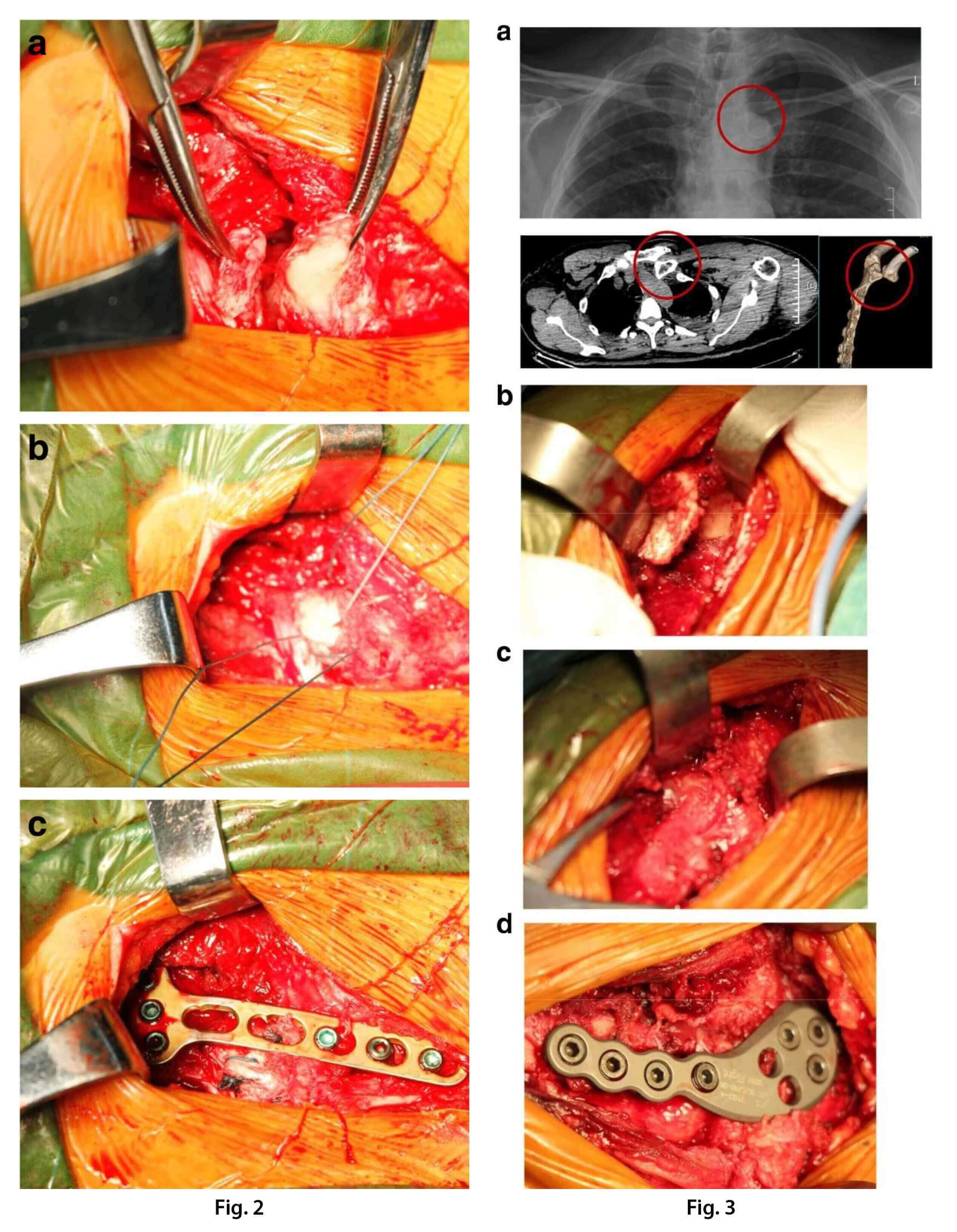
Surgery took place with the patient under general anesthesia and in the supine position. A transverse incision was made at the medial 1/3 of the clavicle and sternum. The skin and subcutaneous tissues were dissected and, if possible, the platysma was incised and raised as a separate layer. The periosteum of the clavicle was reflected superiorly and inferiorly, at which point the affected ligaments of the SC joint could be identified.
In the case of anterior dislocations, it was found that the ligaments and anterior capsule were torn. After manual reduction of the SCJ, the medial end of the clavicle would shift anteriorly, indicating instability. Therefore, SC reduction was maintained, and non-absorbable sutures were used to repair the torn ligaments and capsule, ultimately applying the locking plate for fixation. The plate was placed on the anterior side of the sternum and the medial clavicle. Initial fixation of the medial clavicle was performed using a short drill bit to protect the vascular structures (subclavian artery and vein). Locking screws were used to maintain the SC reduction. Then, the anterior portion of the sternum was carefully drilled, and unicortical locking screws were placed.
For posterior dislocations, the anterior ligament and capsule were dissected to expose the articular surface of the clavicle on the sternal side. Since the position of the medial clavicle was not visible, the clavicle was identified from the lateral side and then followed to the medial end of the clavicle. The affected upper limb was retracted with the shoulder abducted to approximately 90 degrees. The incision was then carefully maintained with forceps. A locking plate was used for fixation in the same manner as for the anterior dislocation. Non-absorbable suture was used to repair the ligament and capsule.
After surgery, the affected limb was immobilized in a brace for 3 weeks, during which passive exercises for the elbow and shoulder were encouraged. Three weeks after surgery, when passive shoulder mobility increased, a gradual transition to active exercises was possible.
All patients were asked to come for a monthly check-up for 3 months after surgery and then every 3 to 6 months after implant removal. Functional assessment was carried out using the Constant Shoulder Score (CSS) questionnaire and the Disability of the Arm, Shoulder and Hand (DASH) questionnaire. Documented post-surgical complications included infection, implant failure, and recurrent dislocation. All plates were removed after 6 months to avoid plate and screw breakage or migration.
Average follow-up was 14 months (range, 11-16 months). Average age, gender distribution, type of dislocation, and associated injuries were tracked.
All patients had secondary surgeries for plate removal at 6 months postoperatively. At final follow-up, the average CSS and DASH scores were 89.5 (range, 78-98) and 9.0 (range, 4-16), respectively.
There were no early complications, such as wound infection or neurological or vascular deficit; screws and plates also did not break or migrate. No cases of redislocation or arthrosis were observed. Four patients were satisfied with the results. All patients were able to return to their previous activities.
Only patient 3 had an unsatisfactory outcome. She had a right clavicular disjunction and a left proximal humeral fracture. Closed reduction for the anterior SC dislocation failed, and open reduction and internal fixation for the two injuries were performed the next day. Postoperative X-ray showed that the right acromioclavicular joint (ACJ) was dislocated - that is, Rockwood type V (Fig. 5). After reviewing the initial CT scan, it was found that the posterior ACJ dislocation was actually a Rockwood type IV. Therefore, a second surgery was performed to treat the clavicular disjunction. The screws previously placed in the clavicle were removed, and reduction of both dislocations was performed simultaneously. A hook plate was used to treat the ACJ dislocation. Three months after the second surgery, the two implants used to treat the disjunction were removed. At the last check-up, although there was no pain or redislocation, abduction of the affected shoulder was limited to 110 degrees.
The SC joint is a saddle-type joint that represents less than half of the medial clavicle, since it articulates with the superior angle of the sternum. When the shoulder girdle moves, the SC joint has a range of motion in three planes. Despite its intrinsic instability due to its bony anatomy, strong soft tissues stabilize the structures - including the ligaments, subclavius muscle, articular disc, and capsule - so that the SC joint is one of the least commonly dislocated joints in the body.
Indirect force applied to the shoulder girdle is the common mechanism of SC dislocation. Occasionally, a direct force applied to the medial clavicle will lead to a posterior dislocation. When medial clavicle prominence is noted after trauma, anterior SCJ dislocation should be considered; a palpable defect at the medial end of the clavicle is the obvious sign of a posterior SCJ dislocation.
After such a diagnosis, it is important that the entire clavicle and the AC joint be examined. Especially for anterior SC dislocation, the acromioclavicular joint should be carefully checked to determine whether or not there is a posterior dislocation. In the case of patient 3, examination of the ipsilateral AC joint was neglected, and the diagnosis of clavicular disjunction was delayed.
.jpg)
Postoperative - A postoperative three-dimensional CT scan a) showing good reduction and fixation with the locking plate, sagittal and transverse CT scans b) showing unicortical screw fixation in the sternum.
.jpg)
A postoperative X-ray showing dislocation of the right acromioclavicular joint
Since it can be obscured by neighboring structures, SC dislocation can easily be missed on X-ray. A CT scan is the best imaging diagnostic method and can help distinguish between medial clavicular fractures and SC dislocations. Careful examination of a three-dimensional CT scan can help determine whether SC displacement is present and whether or not there is also a posterior acromioclavicular dislocation.
Closed reduction is the primary treatment choice for SC dislocation. For anterior traumatic SC dislocations, although there is a risk of recurrent instability, functional deficits rarely occur after closed reduction. However, if the medial area of the clavicle is prominent, surgical correction may be preferred. For posterior traumatic SC dislocations, there may be several potential complications, so such injuries must be treated immediately with closed reduction. When posterior SC dislocation cannot be reduced this way, open reduction must be performed urgently to minimize the risk of cardiovascular compromise.
The goal of the surgery is to restore SC bony anatomy and joint stability. The intraoperative risk is that of damaging neighboring structures at the sternum and medial clavicle level, and also that of causing postoperative migration of the implants. The optimal surgical process should minimize these risks as much as possible.
There are many surgical methods for treating SC dislocation, including ligament repair with reconstruction, fixation with Kirschner wires or pins, as well as plate fixation. The best choice among the different procedures is also controversial. Ligament repair with reconstruction is the most common method according to the literature. These procedures require relatively complex operative manipulation, greater soft tissue dissection, and a prolonged postoperative immobilization time. Fixation with Kirschner wires is contraindicated due to the high risk associated with their migration into vital structures.
To date, three studies have been reported regarding the treatment of SC dislocation using fixation plates, including that of Franck et al., who used Balser plates to treat three posterior SC dislocations. Shuler and Pappas used dual perpendicular locking plates to fix two posterior dislocations, and Hecox et al. used a rim-plating technique to treat two posterior dislocations. The Balser plate requires a hook insertion into the sternum, which appears to endanger vital structures. Dual locking plates can achieve rigid fixation for the SCJ, but due to the additional soft tissue manipulation, the medical cost will be higher. The rim-plating technique, which does not require the use of a drill or screws in the sternum, can clearly avoid damage to vital structures, but the stability of SCJ fixation may be insufficient. The biomechanics involved in this innovative method require further research.
The use of a single locking plate may be a preferable alternative for treating SC dislocations. The purpose of the plate is to maintain the SC reduction and allow the soft tissues around the joint to heal. In the surgical procedure, unicortical screws in the sternum and bicortical screws in the medial clavicle were used to maintain SC stability. Meanwhile, the affected ligaments and capsule were sutured to provide preliminary stability to the joint. After stable fixation of the locking plate, the soft tissues can heal, and the SC joint will be stable. To avoid iatrogenic injuries, only the anterior manubrium of the sternum was drilled, and bicortical screws were used for fixation. Ultimately, the locking plate allows relatively stable fixation, as well as a certain degree of movement. Thus, by using a single locking plate to treat SC dislocation, we can also ensure a certain degree of joint movement, which facilitates healing and recovery of shoulder function.
We believe that fixation with a single locking plate, combined with ligament and capsular repair, is easier to perform, minimizes soft tissue manipulation, and protects the periosteal blood supply. Unicortical drilling and screw fixation in the sternum is a relatively safe surgical process in terms of protecting vital structures. Most of these patients have good function without recurrent dislocation or subluxation.
To avoid migration of loose or broken implants, locking plates must be removed at approximately 3 months, maximum 6 after surgery; this is the disadvantage of this technique.
In addition, this study has some limitations. It is a retrospective study that involved a small number of patients with a short-term follow-up. Long-term follow-up would be necessary to determine whether postoperative arthritis developed in these patients.
Fixation using a single locking plate combined with ligament and capsular repair is relatively easy to perform, decreases the risk of soft tissue injury, and protects the periosteal blood supply. The study indicates that open reduction and fixation with locking plates for the treatment of traumatic sternoclavicular joint dislocations is a safe, relatively simple surgical procedure that can lead to satisfactory results.
Centrokinetic is the place where you will find answers and clear solutions for your movement problems. The clinic dedicated to osteoarticular conditions is divided into the following specialized departments:
Find out the latest news by following the Facebook and YouTube accounts of Centrokinetic clinic
See details
READ MORESee details
READ MORESee details
READ MORESee here how you can make an appointment and the location of our clinics.








CENTROKINETIC FLOREASCA
Bucharest, 1st District,
Mircea Eliade Street, no. 18,
Eliade Tower, Entrance A, Floor 3
| Navigation | Explore 3D |
CENTROKINETIC Barbu Vacarescu
Bucharest, 2nd District,
Barbu Vacarescu Street, no. 102,
102 The Address, ground floor.
| Navigation | Explore 3D |
CENTROKINETIC MILITARI
Bucuresti, Sector 6,
Bld. Iuliu Maniu, nr. 15G,
Cladirea PBT, etaj 3.
| Navigation |
CENTROKINETIC CLUJ-NAPOCA
Cluj-Napoca,
Str. Intre Lacuri, nr.1
Intrarea este in spatele cladirii.
| Navigation |
SOCRATES CLINIC TIMISOARA
Timisoara, Calea Martirilor 1989,
nr. 1.
Telefon: 0371 785 374.
| Navigation |
Phone: 0319693
E-mail: programari@centrokinetic.ro
Operating hours:
Monday – Friday: 07:00 – 21:00
Saturday: 09:00 – 14:00
Sunday: closed.
MAKE AN APPOINTMENT