
Synovial Cyst


- What is a synovial cyst?
- What causes a synovial cyst?
- Types of cysts
- Why do synovial cysts appear?
- Symptoms of synovial cysts
- Diagnosing a synovial cyst
- How a synovial cyst is treated
- Medication and non-invasive treatments
- Injections
- Kinesiotherapy / Occupational therapy
- Surgical treatment
What is a synovial cyst?
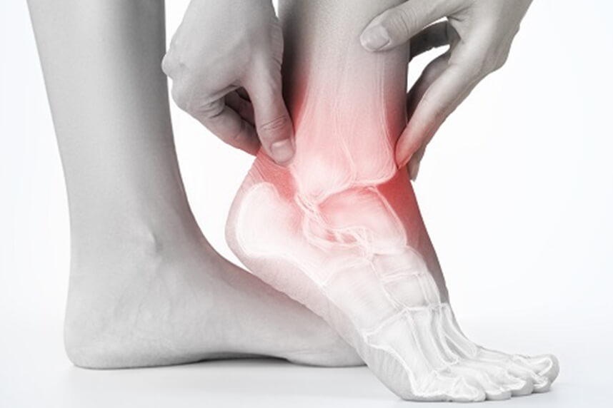
Synovial cysts are non-cancerous formations that most commonly develop along the tendons or joints of the wrists. They are also called ganglion cysts. They can also appear on the ankles and feet. There are also cases of synovial cysts that appear along the spine. Synovial cysts are usually round or oval and are filled with fluid. Small synovial cysts can be the size of a pea, while larger ones can have a diameter of about 2.5 centimeters. Synovial cysts can be painful if they press on a nearby nerve. Their location can sometimes interfere with joint movement.
Synovial cysts that appear along the spine almost always develop in the lower back and are called lumbar synovial cysts. These cysts are less common and are not cancerous. Many synovial cysts will not cause any symptoms. If a lumbar synovial cyst grows large enough, it can lead to spinal stenosis, a condition in which the spinal canal becomes too narrow for the spinal nerves to move freely. This causes pain and cramping in the back and legs, which worsens if the affected person stands for too long.

What causes a synovial cyst?
Synovial fluid, contained within a membrane called the synovial sac, lubricates the joints and helps them move smoothly. As joints age and begin to wear down, cartilage deteriorates. In response, the body may produce more synovial fluid in an attempt to keep the joints moving smoothly. Synovial cysts form when this extra fluid accumulates inside a section of the synovial sac.
Types of synovial cyst
Ganglion cysts of the hand
Ganglion cysts are common in the hand. They are found in common locations and are often seen on the back of the wrist, in the middle. They can also be near a finger joint. A ganglion cyst can occur in patients of all ages. While the cause of ganglion cysts is unknown, cysts can form when the joints or tendons are irritated, in cases of arthritis, mechanical changes, or injuries.
Signs and symptoms of ganglion cysts
Ganglion cysts can appear quickly, can change size, and can disappear without treatment. They are largely harmless, non-cancerous, and most often do not require specialized treatment, only observation.
Symptoms of a ganglion cyst usually begin with a lump appearing under the skin. They can vary in shape and size, some even growing over time. How the cyst affects a person's life depends on the location and size of the cyst. Some patients have no symptoms, while other patients may feel irritation, pain, or tingling if the cysts press on nerves, ligaments, or arteries. Also, as the cyst increases in size, it can make bending the hand a difficult or painful action.
To be sure that the formed mass is a ganglion cyst, doctors can use several diagnostic techniques. The easiest option is transillumination, which involves using light on one part of the body to detect any abnormality. If the light passes through the lump, it is much more likely to contain fluid and be a cyst. In addition, ultrasound or MRI can also be used to diagnose the cyst.
Treatment of ganglion cysts of the hand
Treatment for a ganglion cyst can often be non-surgical. In many cases, these cysts can simply be observed, especially if they are painless. Ganglion cysts may disappear spontaneously. If a cyst becomes painful, limits activity, or otherwise causes problems, several treatment options are available. Different types of non-surgical treatment may include:
- Observation (the patient informs the doctor if changes occur in the ganglion cyst)
- Splints and anti-inflammatory medication, which can reduce pain associated with certain activities
- Aspiration: This is done to remove the fluid from the cyst. Aspiration can be done with or without ultrasound guidance. Recurrence of the cyst is common, since the cyst wall or sac and stalk remain connected to the joint.
If non-surgical options fail to provide relief or if the cyst recurs, surgical alternatives are available. Surgery involves removing the cyst wall, the fluid, and the stalk. It may include removing a part of the joint capsule or the synovial sheath. In the case of ganglion cysts of the wrist, both open and arthroscopic techniques usually give good results. Surgical treatment is generally successful. Even with surgery, the cyst can return, since we do not fully understand the cause and there are no known preventions. If a ganglion cyst recurs and does not hurt, it can be left untreated. If it recurs and is painful, it can be removed again. The doctor will discuss the best treatment options for the patient and provide specific medical advice for their condition.
Synovial cysts of the spine
Facet joints are small stabilizing joints that are located in pairs between and behind each adjacent vertebra at every level of the spine, except for the top two levels. They are weight-bearing structures and are subject to mechanical stress. Facet joints have cartilage and are lubricated with synovial fluid, which is kept in the joint by a capsule that surrounds the joint.
Synovial cysts are fluid-filled sacs that develop in the joints. If large enough, these sacs can put pressure on one or more nerve roots in the spinal canal or where a nerve exits the spinal canal.
Why do synovial cysts appear?
Spinal synovial cysts are generally caused by degenerative changes, such as osteoarthritis, which can develop in a joint over time. Synovial cysts are rare, non-cancerous, and most often develop as a result of wear and tear or joint degeneration. Therefore, they are rarely seen in patients younger than 45 and are most common in those aged 65 or older. Synovial cysts are rarely dangerous. Patients with joint conditions, such as rheumatoid arthritis, are prone to developing a synovial cyst.
Mechanical stress can lead to joint degeneration. As degeneration increases, synovial fluid may accumulate in an attempt to protect the joint. There is an increased risk of cyst formation when there is increased stress and movement at a spinal segment.
With wear, the facet joint cartilage (the elastic material in a joint that provides protection and shock absorption) erodes. In this process, the synovium can form a cyst. Trauma also has degenerative and inflammatory effects on the joints, which can lead to the formation of a synovial cyst.
Most spinal synovial cysts occur in the lumbar region at the L4 or L5 vertebrae. About a third of patients who have a spinal synovial cyst also have a condition known as spondylolisthesis. It is believed that spondylolisthesis causes the formation of synovial cysts due to decreased facet joint stability.
Instability can occur in any area of the spine, but L4-5 is the most common level. If instability occurs, a cyst may develop. However, it is important to note that these cysts can also form without instability.
Symptoms of synovial cysts
The symptoms of a synovial cyst depend on the size and location of the cyst. Some patients with a synovial cyst may have no signs or symptoms. However, some people may suffer from one or more symptoms:
- Severe pain in the lower back
- Difficulty walking or standing
- Numbness or tingling in one or both legs due to spinal stenosis
- Radiating pain in one or both legs, known as sciatica
- Muscle weakness in both legs
- Loss of motor function in the legs
- Cramping, weakness, or a feeling of heaviness with pain in one or both legs, known as neurogenic claudication
- Temporomandibular joint pain and reduced mouth opening in the case of a cyst present in this region
- Rarely, cauda equina syndrome can develop due to the synovial cyst
Diagnosis
Diagnosis of a synovial cyst begins with a complete medical history and physical examination. A synovial cyst is primarily diagnosed by MRI. This allows the cyst to be visualized, and the doctor can observe whether it is compressing a nerve root or causing central stenosis. X-rays, including observation of flexion/extension movements (i.e., bending), are important to rule out excessive movement (i.e., spinal instability) and to look for the presence of degenerative spondylolisthesis. Nerve irritation can occur as a result of these changes.
Electromyography (EMG) is occasionally performed to determine whether there is evidence of nerve damage, rather than just nerve root irritability.
How a synovial cyst is treated
Initial treatments should be non-invasive and include methods such as postural correction, activity modification, and improving muscle flexibility, as well as medication to reduce inflammation and nerve irritability. Such treatments are usually carried out by physiotherapists and coordinated by a specialist physician.
Medication and non-invasive treatments
Nonsteroidal anti-inflammatory drugs (NSAIDs) include common over-the-counter medications such as aspirin, ibuprofen, and naproxen, among others. These medications reduce pain over the long term and do not raise concerns about dependency.
Opioid therapy for chronic back pain control is less ideal due to potential toxicity to the body and physical and psychological dependence. Treatment with this class of medication should generally be a short-term option when patients do not respond to alternatives.
Pain can often be reduced through the use of muscle relaxants, pain medication, antidepressants, and oral steroids.
Injections
In some cases, the doctor may administer a corticosteroid injection to relieve pain and reduce inflammation. Corticosteroids mimic the effects of the hormones cortisone and hydrocortisone, which are produced by the outer layer (cortex) of the adrenal glands. When prescribed in doses exceeding natural levels, corticosteroids suppress inflammation, which in turn relieves pressure and pain. They are most effective injections when used together with a rehabilitation program. In addition, corticosteroids can cause serious side effects, so the number of injections you can receive is limited - usually no more than three in a year.
A steroid is often combined with an anesthetic and injected into the area around the irritated spinal nerves causing the pain. Steroids reduce nerve irritation by inhibiting the production of proteins that cause inflammation. The anesthetic blocks nerve conduction in the area where it is applied, numbing the sensation of pain.
If injections do not provide sufficient pain relief or a return to an acceptable level of functioning, then surgery may be considered.
Kinesiotherapy / Occupational therapy
Physical and occupational therapy can play a vital role in recovery. Once acute pain improves, the doctor or a therapist can design a rehabilitation program to help prevent recurring injuries.
These programs often include heat, cold, and electrotherapy to help ease pain, reduce swelling, increase strength, and promote healing. Methods include therapeutic exercises, manual therapy, functional training, and the use of assistive devices and adaptive equipment to increase strength, range of motion, endurance, speed up wound healing, and functional independence.
Almost all types of treatment will also include some form of exercise. For aerobic exercise, it may be preferable for the patient to try riding a stationary bike instead of walking, since in the seated position (leaning forward) the patient should feel fairly comfortable.
Surgical treatment
Synovial cysts can be removed using an arthroscope. Removing these cysts using minimally invasive techniques minimizes soft tissue trauma and blood loss, and will also result in a smaller incision. In cases where synovial cysts are associated with spondylolisthesis, this technique can minimize the risk of progressive instability and the need for fusion.
The goal of the operation is to remove the cyst to provide more space for the spine to relieve pressure on the nerve roots. The operation is similar to a microdiscectomy. It is not a difficult surgical procedure, but the patient will need time to recover.
To prevent the cyst from reforming, especially in cases where there is associated spondylolisthesis, the surgeon may opt to fuse the affected joint. This type of surgery is called lumbar fusion.
Centrokinetic is the place where you will find clear answers and effective solutions for musculoskeletal conditions. Our clinic, dedicated to musculoskeletal disorders, is organized into the following specialized departments:
- Orthopedics, a department composed of a highly experienced team of orthopedic surgeons specializing in sports traumatology.
- Pediatric Orthopedics, where sports injuries in children (ligament and meniscus injuries), spinal deformities (scoliosis, kyphosis, hyperlordosis), and foot deformities (hallux valgus, hallux rigidus, equinovarus foot, flatfoot, cavus foot) are treated.
- Neurology, featuring a state-of-the-art department where consultations, electroencephalograms (EEG), and electromyography (EMG) examinations are performed.
- Medical Rehabilitation for adults and children, a department specialized in the rehabilitation of professional athletes, spinal disorders, and children with neurological and traumatic conditions. Our extensive experience includes treating more than 5,000 professional athletes.
- Medical Imaging, with the clinic equipped with ultrasound and MRI systems dedicated to musculoskeletal conditions, complemented by an experienced radiologist, Dr. Cosmin Pantu, specialized in musculoskeletal imaging.
- Rheumatology, a comprehensive department dedicated to the diagnosis, treatment, and rehabilitation of patients with non-surgical musculoskeletal disorders.
- Vascular Surgery, a highly specialized department focused on the diagnosis and treatment of vascular diseases affecting arteries, veins, and lymphatic vessels.
- Psychology and Speech Therapy. Neurological and musculoskeletal conditions can have a psychological impact on patients, which is why we believe complete recovery requires addressing both the physical condition and its psychological consequences.
- Neurofeedback. This innovative method helps improve concentration, reduce anxiety, and achieve emotional balance through a simple and interactive process.
Stay up to date by following Centrokinetic on Facebook, Instagram, and YouTube.
MAKE AN APPOINTMENT
FOR AN EXAMINATION
See here how you can make an appointment and the location of our clinics.
MAKE AN APPOINTMENT
BUCHAREST TEAM


































































CLUJ NAPOCA TEAM


































BRASOV TEAM


















Asiguratori privati





Parteneriate



CENTROKINETIC FLOREASCA
Bucharest, 1st District,
Mircea Eliade Street, no. 18,
Eliade Tower, Entrance A, Floor 3

| Navigation | Explore 3D |
CENTROKINETIC Barbu Vacarescu
Bucharest, 2nd District,
Barbu Vacarescu Street, no. 102,
102 The Address, ground floor.

| Navigation | Explore 3D |
CENTROKINETIC MILITARI
Bucuresti, Sector 6,
Bld. Iuliu Maniu, nr. 15G,
Cladirea PBT, etaj 3.

| Navigation |



CENTROKINETIC CLUJ-NAPOCA
Cluj-Napoca,
Str. Intre Lacuri, nr.1
Intrarea este in spatele cladirii.

| Navigation |


SOCRATES CLINIC TIMISOARA
Timisoara, Calea Martirilor 1989,
nr. 1.
Telefon: 0371 785 374.

| Navigation |
CONTACT
Phone: 0319693
E-mail: programari@centrokinetic.ro
Operating hours:
Monday – Friday: 07:00 – 21:00
Saturday: 09:00 – 14:00
Sunday: closed.
MAKE AN APPOINTMENT

