
Testicular Ultrasound: Symptoms That Require Prompt Investigation


 Testicular Ultrasound: Symptoms That Require Prompt Investigation
Pain or a change in the testicle can take you by surprise. Many active men tend to postpone the consultation hoping that the discomfort will go away on its own. In medical practice, this delay can complicate things.
The testicular ultrasound is the first investigation recommended in most scrotal conditions. The procedure is quick, painless, and provides clear information about the structure and vascularization of the testicles. Here is what is important to know about this investigation.
What is a testicular ultrasound and how does it help you?
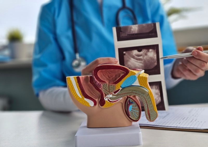
The testicular (scrotal) ultrasound uses ultrasound waves to visualize the testicles, the epididymis, and the spermatic cord. The procedure does not use radiation and requires no special preparation. The doctor applies a gel to the skin and moves the probe to obtain real-time images.
The Doppler variant also allows the evaluation of blood flow. This component makes the difference in situations such as testicular torsion, where circulation is reduced or absent. According to studies, the ultrasound has a high sensitivity for detecting testicular tumors and can differentiate solid lesions from cystic ones. At Centrokinetic you can access complete ultrasound services through the imaging department.
The major benefit of a correct evaluation lies in quickly establishing the diagnosis and choosing the appropriate treatment. Early intervention increases the chances of recovery and reduces the risk of complications. Here are the situations in which a testicular ultrasound may be recommended.
Sudden and intense testicular pain 
Sudden, severe pain in a single testicle is a medical emergency. The most common cause is testicular torsion – the twisting of the spermatic cord which cuts off blood circulation. Frequently associated signs:
- sudden onset of pain;
- nausea or vomiting;
- a testicle positioned higher than the other;
- local swelling and redness.
Surgery performed within the first 6 hours considerably increases the chances of saving the testicle. The Doppler ultrasound shows the lack of blood flow or the specific image of the twisted spermatic cord. Do not wait to see if the pain subsides. Go directly to a urological consultation!
Rapid swelling of the scrotum after trauma
Amateur or professional athletes sometimes suffer blows to the groin area. An impact during a football match or a martial arts training session can lead to a hematoma or even a testicular rupture. The ultrasound can reveal:
- blood accumulation inside the testicle;
- discontinuity of the capsule (tunica albuginea);
- significant tissue edema.
In sports practice, doctors recommend immediate imaging evaluation if the swelling increases rapidly or the pain persists for more than a few hours. Early intervention can prevent the loss of testicular function.
Pain associated with fever
Progressive pain, accompanied by fever and a burning sensation when urinating, suggests an infection such as epididymitis or orchitis. The epididymis is the tubular structure behind the testicle where sperm cells mature.
According to current information, untreated infections can lead to abscesses or impaired fertility. The Doppler ultrasound shows increased blood flow (hyperemia), which differentiates infection from torsion.
The doctor will recommend antibiotic treatment adapted to the cause. Avoid self-medication. Consult the specialist for a correct treatment plan and for monitoring!
A nodule or palpable mass in the testicle
A hard, painless nodule discovered during self-examination requires prompt investigation. Testicular cancer frequently occurs between the ages of 15 and 35, but it can also be diagnosed at older ages. Data show that the survival rate is very good when the disease is detected early. The ultrasound differentiates:
- solid intratesticular lesions (with oncological suspicion);
- benign cysts;
- testicular microlithiasis (small calcium deposits).
Not every nodule means cancer. In most cases, the imaging evaluation can clarify the nature of the lesion and reduce the patient's anxiety. However, only the doctor can determine the next steps: blood tests, tumor markers, or surgery. Perform testicular self-examination monthly, especially if you have a history of an undescended testicle or a family history of oncological conditions.
A feeling of heaviness
This description is typical for a varicocele, meaning the dilation of the veins around the testicle. It occurs more frequently on the left side and becomes more pronounced when standing or after intense physical exertion. The Doppler ultrasound identifies:
- veins dilated beyond 2–3 mm;
- venous reflux during the Valsalva maneuver.
Studies show an association between varicocele and reduced sperm quality. For athletes who perform sustained physical effort or for men planning a pregnancy with their partner, a correct evaluation helps establish a personalized strategy: monitoring or surgical correction.
Progressive increase in scrotal volume without pain
A slow and painless enlargement can indicate:
- hydrocele (fluid around the testicle);
- spermatocele (cyst of the epididymis);
- epididymal cyst.
The ultrasound makes a clear distinction between fluid and solid mass, similar to a soft tissue ultrasound. In many situations, the hydrocele does not require immediate intervention and can be monitored. The doctor decides based on size, discomfort, and functional impact.
Testicular pain in a child or adolescent
Parents must treat any testicular pain in a child with the utmost attention. Testicular torsion frequently occurs in adolescents, and the intervention window is limited.
The testicular ultrasound is safe at any age and does not use radiation. Do not attribute the pain to a minor blow without a medical evaluation. A prompt consultation can prevent long-term complications.
What other conditions can the testicular ultrasound identify?
In addition to the situations above, the investigation can detect:
- an inguinal hernia descended into the scrotum;
- repeated minor traumas;
- changes associated with infertility;
- vascular lesions.
For a complete evaluation of the male urogenital system, the doctor may also recommend a penile Doppler ultrasound.
In certain cases, patients recovering after pelvic procedures may present associated urinary symptoms. Dedicated urinary incontinence recovery programs can complement medical treatment.
How is the investigation performed?
The procedure takes approximately 15–20 minutes. You lie on your back, and the doctor applies a transparent gel to the examined area. The ultrasound probe transmits ultrasound waves and displays the images on the monitor. You do not need:
- fasting;
- a full bladder;
- to interrupt your usual treatments (the decision belongs to the doctor).
The investigation does not cause pain. You can resume your activities immediately after it is completed. Similar to the musculoskeletal ultrasound used in orthopedic pathology, the method can be repeated for monitoring.
The difference between the classic ultrasound and the Doppler ultrasound
| Characteristic | Simple testicular ultrasound | Doppler ultrasound |
|---|---|---|
| What does it visualize? | The anatomical structure, shape, and size of the testicle. | Blood flow and the direction of blood circulation. |
| Main use | Identifying nodules, cysts, or fluid (hydrocele). | Diagnosing torsion (absent flow) or infection (increased flow). |
| In trauma | Detects tissue ruptures or hematomas. | Checks whether the testicle is still supplied with blood after the blow. |
| In varicocele | Measures the diameter of the dilated veins. | Confirms blood reflux in the veins when you exert effort. |
Why does early intervention matter?
An evaluation performed in time offers several benefits:
- it establishes the correct diagnosis;
- it reduces the risk of complications;
- it allows the appropriate treatment to be chosen;
- it increases the chances of preserving testicular function.
In urological practice, the difference between a problem treated promptly and one that is ignored can influence fertility, comfort, and physical performance. The medical team analyzes each case individually and establishes a personalized plan, adapted to your age, activity level, and medical history.
If you notice recent changes or persistent symptoms, do not postpone the consultation. Book an appointment at Centrokinetic now for a specialist consultation!
Frequently asked questions
Is the testicular ultrasound painful?
No. The investigation is painless and does not require anesthesia. You may feel slight pressure from the probe on the sensitive area, but the discomfort is minimal and short-lived.
How quickly should I have the ultrasound if I have sudden pain?
Immediately. Intense pain that appears suddenly can indicate testicular torsion, a situation that requires prompt intervention. Go to the emergency room or to an urgent urological consultation.
Can I have the ultrasound if I have no symptoms, just for a check-up?
In the absence of symptoms, the ultrasound is not routinely recommended for all men. The doctor may indicate it if you have risk factors, a history of varicocele, an undescended testicle, or changes discovered during a clinical examination. Talk to the specialist for a recommendation adapted to your situation.
Disclaimer: This material is for informational purposes and does not replace a specialist medical consultation. In case of severe pain that appears suddenly, seek urgent medical attention!
Sources:
- "Testicular Ultrasound" Cleveland Clinic, 13 June 2024. Accessed 16 Apr. 2026.
- Mariz, Jose, et al. "Testicular Ultrasound: An Emergency Medicine Perspective." Internal and Emergency Medicine, vol. 20, no. 4, 18 Mar. 2025, pp. 1153–1163, 10.1007/s11739-025-03864-z. Accessed 16 Apr. 2026.
BUCHAREST TEAM


































































CLUJ NAPOCA TEAM


































BRASOV TEAM


















MAKE AN APPOINTMENT
FOR AN EXAMINATION
See here how you can make an appointment and the location of our clinics.
MAKE AN APPOINTMENT
Asiguratori privati





Parteneriate



CENTROKINETIC FLOREASCA
Bucharest, 1st District,
Mircea Eliade Street, no. 18,
Eliade Tower, Entrance A, Floor 3

| Navigation | Explore 3D |
CENTROKINETIC Barbu Vacarescu
Bucharest, 2nd District,
Barbu Vacarescu Street, no. 102,
102 The Address, ground floor.

| Navigation | Explore 3D |
CENTROKINETIC MILITARI
Bucuresti, Sector 6,
Bld. Iuliu Maniu, nr. 15G,
Cladirea PBT, etaj 3.

| Navigation |



CENTROKINETIC CLUJ-NAPOCA
Cluj-Napoca,
Str. Intre Lacuri, nr.1
Intrarea este in spatele cladirii.

| Navigation |


SOCRATES CLINIC TIMISOARA
Timisoara, Calea Martirilor 1989,
nr. 1.
Telefon: 0371 785 374.

| Navigation |
CONTACT
Phone: 0319693
E-mail: programari@centrokinetic.ro
Operating hours:
Monday – Friday: 07:00 – 21:00
Saturday: 09:00 – 14:00
Sunday: closed.
MAKE AN APPOINTMENT

