See details
READ MORE

.jpg)
This article was written by dr. Aursulesei Ruxandra, pediatric neurologist with extensive experience in diagnosing and treating neurological conditions. Pediatric neurology plays an important role in the periodic neurological evaluation of children born prematurely, as well as of all newborns at risk of developing neurological sequelae, with the goal of early detection and treatment of any impairment of the nervous system.
The Kleeblattschädel anomaly is the most severe form of craniosynostosis, caused by intrauterine synostosis of all the sutures.
The skull has a "cloverleaf" appearance.
Craniofacial changes include:
Children present with hydrocephalus, psychomotor retardation, blindness, obstruction of the nasolacrimal duct, absence of the external auditory canal – deafness, and corneal ulcerations.
Brachycephaly forms through the premature closure of both coronal sutures. It occurs in approximately 20% of craniosynostosis cases and is more commonly found in females.
The skull is flattened in the antero-posterior direction, with the bitemporal diameter enlarged compared to normal. The occiput and forehead are flattened. The anterior fontanelle is positioned much further forward, and a bony "ridge" can sometimes be palpated at the level of the prematurely closed coronal sutures.
Children may present with strabismus, papilledema, or optic atrophy.
Skull X-ray may show an elliptical orbit or the harlequin eye sign.
.jpg)
.jpg)
.jpg)
Oxycephaly forms as a result of the premature closure of all the sutures.
The skull is excessively tall and narrow, with the anterior fontanelle remaining open, allowing the skull to grow only at this level, taking on a pointed, "cone" shape.
This type of craniosynostosis can cause a marked increase in intracranial pressure and significant neurological sequelae.
It can cause: optic atrophy, divergent strabismus, papilledema, exophthalmos, choanal atresia, anosmia, bilateral pyramidal syndrome, global developmental disorder, cognitive-behavioral disorders, Chiari I malformation, papilledema.
Hearing and vestibular disorders may occur due to narrowing of the internal auditory canal.
.jpg)
Plagiocephaly is a form of craniosynostosis that usually occurs through the premature fusion of one coronal suture or, more rarely, of the lambdoid sutures unilaterally.
The skull is asymmetric, and facial deformities may also be present.
In unilateral synostosis of the lambdoid suture, the ipsilateral ear is displaced forward, one frontal bone is larger than the other, and the contralateral occiput is larger and flat. The shape of the head resembles that of a parallelogram.
.jpg)
.jpg)
Plagiocephaly caused by synostosis of one coronal suture
Scaphocephaly represents the premature closure of the sagittal suture. This is the most commonly encountered craniosynostosis (60% of all craniosynostosis cases).
The antero-posterior diameter of the skull is greatly enlarged, narrowed transversally, the forehead bulges, and a bony ridge can be palpated at the level of the fused sagittal suture (sometimes even from birth).
Neurological examination is normal in most cases.
.jpg)
.jpg)
.jpg)
Chotzen syndrome is a condition characterized by unilateral/bilateral coronal craniosynostosis.
The face is asymmetric, with a deep (ogival) palate; patients may have dental crowding, supernumerary teeth, and enamel hypoplasia. It is also associated with brachydactyly, syndactyly, and various skeletal abnormalities.
Chotzen syndrome can be associated with developmental disorders, learning difficulties, mental retardation, speech disorders, cryptorchidism, hearing disorders, strabismus, cleft palate, optic atrophy, cardiac malformations, and renal malformations.
Apert syndrome is an autosomal dominant disease that occurs through mutation of the FGFR2 gene located on chromosome 10.
This syndrome is also known as acrocephalosyndactyly (concurrent malformations of the skull and limbs)
At the cranial level, bilateral synostosis of the coronal sutures occurs, causing turribrachycephaly.
At the facial level, the patient is distinguished by the following clinical signs: flattened forehead, sunken temporal area, proptosis (or exophthalmos), and hypertelorism. The nose is short, wide, and with a sunken base. The nasolabial angle is diminished.
The greater wing of the sphenoid is protruded.
It also presents with orbital hypoplasia and hypertelorism, maxillary hypoplasia, and complete and symmetric syndactyly of the 2nd, 3rd, and 4th fingers.
The lower limbs are also affected, but less severely. The patient's stature is short.
The palate is arched, with an appearance similar to a cleft lip and palate, but with the hard palate remaining intact. This typical cleft-like appearance is given by the presence of lateral swellings at the palatal level.
The uvula can be bifid in approximately 75% of cases.
Class III malocclusions often occur due to the developmental deficit of the maxillary bone mass, associated with anterior open bite. Supernumerary teeth, ectopic eruptions, and milky-white opacities of the enamel may occur. The periodontium (gum) is hypertrophic, with increased thickness.
Middle ear abnormalities may occur, contributing to hearing disorders.
Children diagnosed with Apert syndrome may have mental retardation, hydrocephalus, headaches, Chiari malformation, and epileptic seizures.
Recurrent ear infections occur and can lead to hearing loss, or patients may present with hearing loss from birth, depending on the degree of bone involvement. Additionally, patients may also present with hyperhidrosis (excessive sweating), acne-prone skin, eyebrow alopecia, or fusion of the cervical vertebrae.
Crouzon syndrome is a genetic disease, transmitted in an autosomal dominant manner. It occurs through mutation of the gene encoding FGFR2, rarely FGFR1 (fibroblast growth factor receptor), located on chromosome 10.
Typically, coronal suture synostosis (unilateral or bilateral) occurs at the cranial level, but it can also be associated with synostosis of the lambdoid, sagittal, and metopic sutures.
A small proportion of patients with this syndrome may present with subluxations of the radial head or minor hand abnormalities. The nose has a beak-like appearance, and the tongue is large.
These children present with exophthalmos, strabismus, hypertelorism, hypoplasia of the maxilla and zygomatic bone
Epileptic seizures occur in 12% of patients. Cervical spine abnormalities can be found in approximately 30%.
It is rarely associated with hydrocephalus. Mental deficits are found in only 3% of patients.
Trigonocephaly is the consequence of the premature closure of the metopic suture.
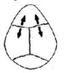
Children have a pointed forehead with a prominent ridge in the middle area of the forehead. It is narrow, constantly accompanied by hypotelorism and the disappearance/flattening of the frontal bosses, so that, when viewed from above, the skull appears triangular.
Some patients may have mental retardation, cleft palate, coloboma, ocular abnormalities, holoprosencephaly, urinary tract abnormalities, Chiari I.
Turricephaly represents synostosis of the sagittal suture and the coronal sutures.
The clinical appearance is that of a tower skull.
Turricephaly is most often associated with intracranial hypertension, global developmental disorder, cognitive-behavioral disorders, Chiari I malformation, or papilledema.
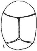


Centrokinetic is well prepared for this specialty, with a team of experienced doctors and the necessary equipment for diagnosis, monitoring, and treatment. Find out more about pediatric neurology and book an appointment at Centrokinetic here.
See details
READ MORESee details
READ MORESee details
READ MORESee here how you can make an appointment and the location of our clinics.








CENTROKINETIC FLOREASCA
Bucharest, 1st District,
Mircea Eliade Street, no. 18,
Eliade Tower, Entrance A, Floor 3
| Navigation | Explore 3D |
CENTROKINETIC Barbu Vacarescu
Bucharest, 2nd District,
Barbu Vacarescu Street, no. 102,
102 The Address, ground floor.
| Navigation | Explore 3D |
CENTROKINETIC MILITARI
Bucuresti, Sector 6,
Bld. Iuliu Maniu, nr. 15G,
Cladirea PBT, etaj 3.
| Navigation |
CENTROKINETIC CLUJ-NAPOCA
Cluj-Napoca,
Str. Intre Lacuri, nr.1
Intrarea este in spatele cladirii.
| Navigation |
SOCRATES CLINIC TIMISOARA
Timisoara, Calea Martirilor 1989,
nr. 1.
Telefon: 0371 785 374.
| Navigation |
Phone: 0319693
E-mail: programari@centrokinetic.ro
Operating hours:
Monday – Friday: 07:00 – 21:00
Saturday: 09:00 – 14:00
Sunday: closed.
MAKE AN APPOINTMENT