What is a renal Doppler ultrasound?
Renal Doppler ultrasound (sometimes simply called "renal ultrasound") is an evaluation that uses ultrasound to provide precise images of blood vessels, with a focus on analyzing their structure and size. Through Doppler ultrasound, blood flow (direction, speed, volume) can be measured both in arteries and veins. This principle applies not only to carotid artery ultrasound (also known as carotid Doppler), but also to vascular Doppler ultrasounds of the lower or upper limbs, as well as cardiac Doppler ultrasound.
When is a Doppler ultrasound of the renal arteries recommended?
There are situations in which the renal arteries (the vessels that carry blood to the kidneys) are narrowed. This narrowing (also known as stenosis) can be congenital (from birth), or acquired as a result of aging, diabetes mellitus, or high cholesterol. In such cases, the patient may have high blood pressure at a young age, difficult to control with medication, or may develop kidney disease. This type of ultrasound can determine if the blood supply to the urinary system is adequate and can confirm or rule out whether issues are caused by low renal blood flow. Renal artery Doppler ultrasound is also recommended in the following situations:
- The patient complains of back pain, which the doctor suspects may be related to a kidney condition;
- Electrolyte imbalances found in routine blood tests – the most common are hyperkalemia (blood potassium levels above normal), hyperphosphatemia (blood phosphate ion levels above normal), hypocalcemia, accompanied by metabolic acidosis.
What happens during this investigation?
The patient must undress to the waist and lie on their back. A probe will be used by the doctor on the patient's abdomen (belly) with a special water-based gel that helps eliminate air between the probe and the skin so that ultrasound waves can transmit more effectively through the skin. The probe is connected via a cable to a machine that processes the ultrasound signals and displays images on a screen. Pulses of ultrasound (sound waves with frequencies too high to be heard by the human ear) are sent through the probe into the skin and down to deep structures, then return as echoes and are recorded by the ultrasound machine in real time. The images are then compiled to show not only the structure but also the movement of vessels (wall pulsations and blood circulation). The waves are reflected like echoes by the various tissues they encounter and then return to the transducer. It analyzes the reflected waves, which travel at different speeds depending on the type of tissue they encounter. Thus, ultrasound travels fastest through bone tissue and slowest through air. The doctor moves the probe over the patient's abdomen, pressing in some areas to obtain the clearest images. Unlike standard abdominal ultrasound, during Doppler ultrasound, certain sound waves obtained can also be heard by the human ear through analysis software. Immediately after the imaging investigation, the patient can resume both physical activities and diet as usual, with no restrictions.
How should the patient prepare for the evaluation?
The renal arteries are located deep in the abdomen, so imaging them can be quite difficult. To facilitate the transmission of ultrasound waves to the level where the renal arteries are located, the patient must follow special preparation for 3 days prior to the investigation:
- walking for at least 1 hour daily is recommended;
- do not use laxatives, Triferment, or activated charcoal;
- avoid foods that cause bloating (raw fruits and vegetables, cabbage, beans, fried foods), and avoid drinking mineral water;
- the evening before the examination, drink unsweetened tea and eat a slice of toast;
- do not eat or chew gum on the day of the examination until the ultrasound is completed.
How long does the examination take?
Renal artery Doppler ultrasound can take about 20 minutes, but sometimes longer depending on image clarity and the number of special measurements required. If abnormalities are found, such as narrowings, atheromatous plaques, or blood clots, it is very important for the doctor to carefully evaluate all characteristics of the identified lesion. In this way, the ultrasound report will include a detailed and accurate description, based on which an effective treatment plan tailored to the patient's needs can be developed.
Can any risks occur after this procedure?
Both standard ultrasound and Doppler ultrasound are types of investigations that pose no risk to the patient. A Doppler ultrasound is completely non-invasive, painless, and does not involve any radiation exposure, unlike X-rays or CT scans that require the use of X-rays. Ultrasound is an extremely effective and fast method through which the health status of the renal arteries can be quickly evaluated. A sensation of pressure or slight discomfort may occur when the probe touches more sensitive areas. However, this feeling usually disappears shortly after the examination is completed. It is important to know that a renal artery ultrasound poses no risk to the patient and can be safely repeated as many times as needed. Another major benefit of ultrasound is that it does not involve the use of contrast substances, unlike other imaging methods for evaluating the renal arterial system, such as angiography or angioCT. This is a very useful advantage for patients who have allergies to contrast substances.
What diagnoses can be made based on this investigation?
With the help of kidney ultrasound, a diagnosis of renal artery stenosis can be made, which means a narrowing of the artery that supplies blood to the kidney. The two most important mechanisms of renal artery stenosis are:
- The process of atherosclerosis (buildup of atheromatous plaques), which particularly affects the proximal segment of the renal artery—where it branches off from the aorta on its way to the kidney. Atherosclerosis in the renal arteries is a sign that raises suspicion of simultaneous involvement of peripheral arteries in other locations. Therefore, such a finding should be followed by further investigations to assess the condition of other important peripheral or central arteries in the body.
- Fibromuscular dysplasia, a condition that affects either the distal portion of the renal artery or even its intrarenal segments.
Doppler Ultrasound Prices
You can find a detailed list of individual service prices here. However, any proper healing process is based on a mixed plan of therapies and procedures, personalized depending on the condition, stage, patient profile, and other objective medical factors. Therefore, to configure a treatment plan—with the therapies involved, durations, and the related costs—please schedule an initial consultation here. Centrokinetic is the place where you will find clear answers and solutions for motor function issues. The clinic dedicated to musculoskeletal conditions is divided into the following specialized departments:
- Orthopedics – a department composed of a highly experienced team of orthopedic doctors specialized in sports traumatology.
- Pediatric orthopedics – where children’s sports injuries (ligament and meniscus injuries), spinal deformities (scoliosis, kyphosis, hyperlordosis), and foot deformities (hallux valgus, hallux rigidus, clubfoot, flatfoot, cavus foot) are treated.
- Neurology – equipped with a high-performance department where consultations, electroencephalograms (EEG), and electromyographies (EMG) are performed.
- Medical recovery for adults and children – a department specialized in the recovery of professional athletes, spinal conditions, and rehabilitation of children with neurological and traumatic conditions. Our experience is extremely extensive, having treated over 5000 professional athletes.
- Medical imaging – the clinic is equipped with ultrasound and MRI machines dedicated to musculoskeletal conditions, and supported by an experienced radiologist: Dr. Cosmin Pantu, specialized in musculoskeletal imaging.
- Rheumatology – a complete department focused on the diagnosis, treatment, and recovery of patients with non-surgical locomotor system conditions.
- Vascular surgery – a highly specialized department in the diagnosis and treatment of blood vessel diseases, including arteries, veins, and lymphatic vessels.
- Psychology and speech therapy – Neurological or musculoskeletal conditions can have a psychological impact on the patient, which is why we believe full recovery also requires addressing the psychological consequences in addition to the physical issues.
- Neurofeedback – This revolutionary method helps you improve concentration, reduce anxiety, and achieve emotional balance, all through a simple and interactive process.
Stay up to date by following Centrokinetic’s Facebook, Instagram, and YouTube accounts.
MAKE AN APPOINTMENT
FOR AN EXAMINATION
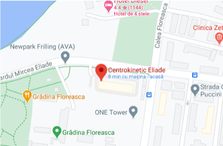
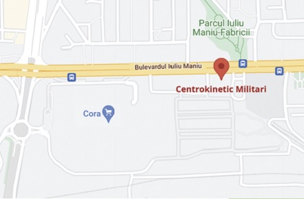
See here how you can make an appointment and the location of our clinics.
MAKE AN APPOINTMENT

.jpg)


