Actualizat: 28-06-2022 / Publicat: 04-07-2017
Recovery protocol in the operated metatarsal V fracture
The forefoot bones play an important role in normal walking. Studies have shown that the two sesamoid bones and the head of the 2-5 metatarsals share body weight evenly while walking.
General principles
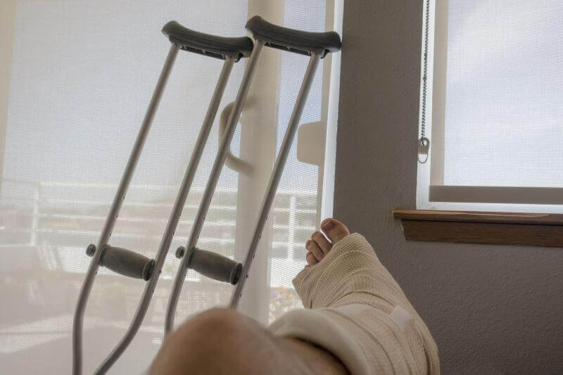
- The patient will use the crutches, without stepping on the operated leg, until the edema is in remission
- Postoperatively, the patient will have his foot immobilized in a walker for 1-2 weeks
- Avoid walking or standing for long periods in the first week to control local edema and pain.
- Apply ice 3-5 times a day (15 minutes each time) to control edema and the inflammatory process
- Raise the operated leg above the level of the heart to limit edema and the inflammatory process
- No impact exercises in the first 6 weeks post-surgery
Causes
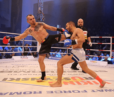
The mechanism of production is usually a sports injury or car accident. Fractures are divided according to location into metatarsal base fractures and distal spiral fractures (dancer's fracture). In addition, the basic metatarsal fractures are divided into 3 areas.
Zone 1: are avulsion fractures, produced by intense internal rotation of the foot with the weight left on the lateral metatarsus, this mechanism putting tension on the insertion of the lateral band of the plantar aponeurosis.
Zone 2: are Jones fractures at the metaphyseal-diaphyseal junction. The most common mechanism is tension on the lateral edge of the metatarsal base.
Zone 3: are the fractures of the proximal diaphysis - they are rarer and found especially in athletes and are located in 1.5 cm distal of the diaphysis. The mechanism of production is the repetitive loading of weight on the lateral metatarsus (jogging).
Zone 1 fractures can be treated orthopedically and symptomatically. Symptoms may persist for up to 6-8 weeks. Zone 2 fractures can be treated orthopedically by plastering and walking with partial or surgical support. Studies have shown that orthopedic treatment for 8-10 weeks is ideal. Zone 3 fractures require more aggressive treatment, which consists of surgery and internal fixation with a screw.
Rehabilitation protocol
Week 1Begin easy ankle mobilization exercises immediately postoperatively
Crochet walking exercises to minimize compensation
Exercises- Active finger movements
- Lower limb flexion with extended knee
- Quadriceps / gluteal muscle exercises
- Cycling
Week 2 - 6
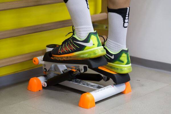
- Start stepping on the operated limb, under the close supervision of the physical therapist, depending on the pain and local edema.
- Radiological examination at 4-6 weeks to monitor healing
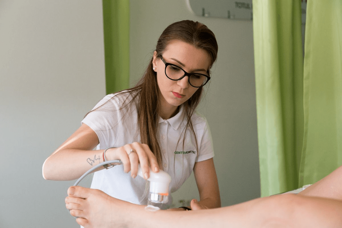
- Soft tissue treatments for local edema prevention, mobility, and soft tissue healing
Exercitii - Cycling, balance exercises
- Stretching in the ankle and foot area and exercises with the help of Theraband bands to regain complete mobility of the foot, as well as to strengthen it
Week 6 - 8 - Progress the functional loading exercises and activities on the operated leg. Avoid jumps and quick changes in direction.
- Continue mobilizing your ankle and foot joints
![]()
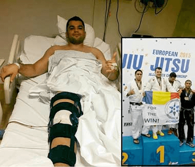
After week 8 - Start the exercises specific to the sport you practice
- Increase the level of strengthening, balance, and coordination training for a gradual return to the specific activities of the sport practiced
- Returning to sports activities depends on a complete functional evaluation, specific to your sport, performed by the physical therapist.