Actualizat: 08-06-2026 / Publicat: 17-10-2018
You can see below more information about MRI:
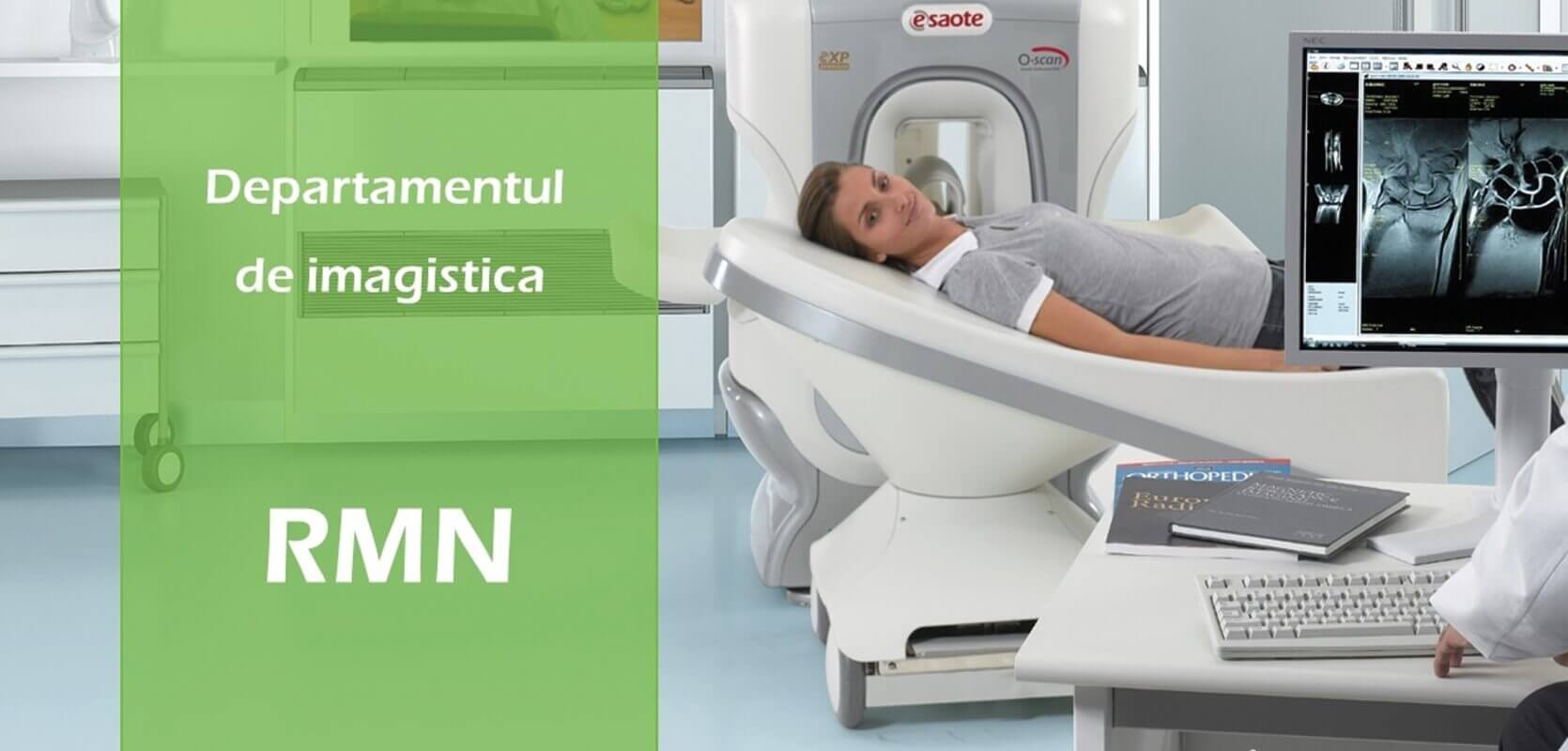
Magnetic resonance imaging (MRI) is a common, non-radiant procedure.
MRI uses a strong magnetic field and radio waves to create detailed images of organs and tissues in the body.
Since its invention, physicians and researchers have continued to refine MRI techniques to assist in medical procedures and research. The development of MRI has revolutionized medicine.
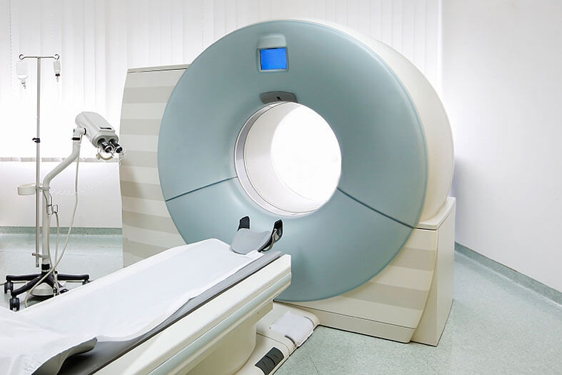
MRI scanning uses a large magnet, radio wave, and a computer to create a detailed, cross-sectional image of internal organs and structures. The scanner typically resembles a large tube with a table in the middle that allows the patient to enter the tube.
An MRI scan differs from CT and X-ray scans because it does not use potentially harmful ionizing radiation.
How safe an MRI is
The strong magnetic field of the NMR system will attract objects that contain iron (also known as ferromagnetic) and can cause them to move suddenly and with great force. This can be a possible risk to the patient. Medical personnel ensures that external objects, such as ferromagnetic screwdrivers and oxygen tanks, are not inserted into the area of the MRI system. As a patient, it is essential to remove all metal objects before the MRI examination, including hearing aids, watches, jewelry, cell phones, and clothing that has metal wires or fasteners. Also, makeup, nail polish, or other cosmetics that may contain metal particles should be removed if applied to the body area, subjected to MRI examination.
The strong magnetic field of the MRI system will pull any object that contains iron in the body, such as certain aneurysm clamps or braces. The MRI technologist and radiologist must be informed of the presence of implants and metal materials so that special precautions can be taken.
The system's magnetic field can damage an external hearing aid or cause a pacemaker, electrical pacemaker, or neurostimulator to malfunction. If you have another metal fragment in your body, there is a risk that it will change position and cause injury.
Also, a metal implant or other object may cause loss of signal or distortion of MRI images.
There are no other hazards or side effects associated with MRI scanning. The test is not painful. Because there is no radiation, the procedure can be repeated without problems. There is a theoretical low risk for the fetus in the first 12 weeks of pregnancy and therefore no scans are performed in pregnant women during this time.
Because patients must be inside a large cylinder while scans are being done, some people become claustrophobic during the test. Patients who are afraid of this should talk to their doctor beforehand, who may give them medication to help them relax.
The device also makes noise while operating, which may be unpleasant.
![i.php?p=aparat-rmn.jpg]()
How long does the investigation take
Usually, an MRI scan takes 30-80 minutes, depending on the examined region and the number of images.
- MRI spine 1 region - scan time 40 minutes;
- MRI spine 2 regions - scan time 60 minutes;
- MRI spine 3 regions - scan time 80 minutes;
- MRI hip / pelvis - scan time 50 minutes;
- MRI knee - scan time 40 minutes;
- MRI ankle - scan time 50 minutes;
- MRI shoulder - scan time 40 minutes;
- MRI elbow - scan time 40 minutes;
- MRI thigh - scan time 40 minutes;
- MRI calf - scan time 40 minutes;
- MRI arm - scan time 40 minutes;
- MRI forearm - scan time 40 minutes;
- MRI hand / wrist - scan time 40 minutes;
- MRI foot / forefoot - scan time 40 minutes;
- MRI sacroiliac joints - scan time 40 minutes;
- MRI sacrococcygeal - scan time 40 minutes;
- MRI midfoot and forefoot - scan time 60 minutes;
- MRI calcaneus - scan time 50 minutes.
MRI scanners use very strong magnetic fields and radio waves, which interact with protons in tissues to create a signal that is then processed to form images of the body. Protons (hydrogen atoms) can be thought of as small magnets with the north and south poles rotating on an axis - like a planet. Normally, protons are arranged randomly, but when a strong magnetic field is applied, the protons are immediately organized.
Applying a radio pulse at the correct frequency stimulates the protons, causing them to resonate and disrupt the magnetic arrangement. Stimulated protons release the absorbed energy as a radio frequency signal and the emission is picked up by a receiving coil in the scanner. The radio frequency that determines the resonance of protons depends on the resistance of the magnetic field. In an MRI scanner, gradient coils are used to vary the intensity of the magnetic field on the body. This means that different sections of the body will resonate at different frequencies. So, by applying sequences of different frequencies, images can be made in thin slices from separate sections of the body. An image is gradually built. When the radio source is turned off, the protons will return to their original state by transmitting radio waves.
Several radio pulse sequences can be used to highlight or suppress certain types of tissues. For example, abnormalities are not usually found in fat, so a sequence of fat suppression pulses can be used to remove the signal from fatty tissue, leaving only the signal in areas with multiple irregularities.
MRI scanners require incredibly strong magnetic fields; generally around 1.5 Tesla, but can also be at 7 Tesla. For comparison, the Earth's magnetic field is only 0.00005 Tesla. The magnet is composed of several coils of conductive wire through which a current passes to generate the magnetic field. To obtain the required intensity, the magnet is cooled with liquid helium below 10 Kelvin (-442oF / -263oC). This allows superconductivity, allowing current to flow through the coils without creating electrical resistance, which means that when a magnet is very cold, it can conduct higher currents and therefore produce stronger magnetic fields.
MRI is widely used in medical diagnosis and, unlike X-rays and CT scans, has the great advantage of not exposing the patient to ionizing radiation. However, the effects of high magnetic fields on the body are still unknown. MRI scanners are especially useful for neurological scanning and are excellent for visualizing small tumors, dementia, epilepsy and other central nervous system disorders.
Scanning table - The patient can be scanned only from inside the magnetic coil so that a motorized table slides him inside and out.
RF system: The antenna produces a radio signal to stimulate the hydrogen nucleus and listen to the radio waves it emits.
Liquid helium: the liquid helium is pumped through a closed shell to cool the superconducting magnets close to absolute zero.
Main magnet: Superconducting magnetic coils produce a magnetic field of 1.5 tesla - about 300 times stronger than a refrigerator magnet.
Gradient system: A second coil distorts the main magnetic field so that the resonant frequency of the protons varies depending on the position.
RMN este procedura preferata pentru diagnosticarea unui numar mare de probleme potentiale, sau a unor afectiuni anormale in multe parti ale corpului. In general, RMN creeaza imagini care pot arata diferente intre tesuturile sanatoase si cele nesanatoase. Medicii folosesc RMN-ul pentru a examina creierul, coloana vertebrala, articulatiile (de ex. genunchi, umar, sold, incheietura mainii si glezna), abdomen, regiunea pelviana, sanul, vasele de sange, inima si alte parti ale corpului. Scopul RMN depinde de ce parte a corpului este investigata.
An MRI of the brain and spinal cord helps the doctor diagnose:
- An aneurysm (bulging or weak blood vessel in the brain)
- Brain tumor
- Brain injuries
- Multiple sclerosis (a disease that affects the outer layer that protects nerve cells)
- Problems with the eye or inner ear
- Spinal cord injuries
- Stroke
A special type of MRI called functional MRI (fRMN) checks the activity of the brain by measuring blood flow to certain areas of the brain. An fRMN can show the active areas of the brain while doing an activity. Your doctor may use this test to see the damage caused by Alzheimer's disease or a brain injury, or before surgery for epilepsy or brain tumors.
An MRI scan of the heart or blood vessels can help diagnose:
- Blockages or swelling in the blood vessels
- Damage caused by a heart attack
- Heart valve problems
- Pericarditis (inflammation of the tissue surrounding the heart)
- Aortic problems (the main artery in the body)
- Problems with the structure of the walls and chambers of the heart
- Tumors inside the chambers of the heart
Brain disorders
MRI can detect a variety of brain conditions, such as cysts, tumors, bleeding, swelling, developmental and structural abnormalities, infections, inflammatory conditions, or problems with blood vessels. It can detect brain damage caused by injury or stroke.
Brain MRI can be helpful in assessing problems such as persistent headaches, dizziness, weakness, and blurred vision or seizures, and can help detect certain chronic diseases of the nervous system, such as multiple sclerosis.
In some cases, MRI can provide clear images of parts of the brain that cannot be seen with an X-ray, CT scan, or ultrasound, making it especially valuable for diagnosing problems with the pituitary gland and brainstem.
Spinal disorders
While MRI scanning is a very sensitive and accurate assessment of spinal anatomy, it cannot distinguish between painful and painless structures of the spine. In fact, a patient may have severe back pain and an MRI scan may show a relatively normal spine or, conversely, the patient may not have back pain, but the MRI scan highlights a lot of anatomical problems. Thus, the conclusions regarding the MRI scans do not constitute a diagnosis, and the MRI results must be correlated with the patient's physical examination and the symptoms of back pain in order to reach a clinical diagnosis.
You may need this scan to check for spinal conditions, including:
- Low back pain
- Cervical pain
- Numbness, tingling, and weakness in the arms and legs
MRI can scan the entire spine or just part of it. MRI can check the discs and nerves of the spine for conditions such as spinal stenosis and spinal tumors.
When a doctor orders an MRI scan for the spine, there is specific information that he is looking for. The information usually obtained from MRI results includes:
- Arrangement of the spine
- Disc height and hydration
- The structure of the vertebral body
- Intervertebral disc - What does the disc look like - If it is normal, clogged, herniated, dehydrated, or degenerated.
- Spinal canal size - If there is enough space for the spinal cord and nerves. If there is compression of the spinal cord or nerve root.
- Nerves - If there are "pinched" (compressed) or inflamed nerves.
- Abnormalities - If there is an abnormality around the spine that could stimulate back pain.
- Spinal cord - If the spinal cord is looking normal
- Postoperative - If anything has changed since the spine operation. If there is another herniated disc, postoperative scarring, or infection.
An MRI of the foot is done to identify the cause of any pain or discomfort in the given area. An MRI is useful because it shows the doctor if there are any abnormalities in the bones or joints. An MRI of the foot will allow the doctor to see the soft tissues of your body besides the bone, which allows him to see all the elements of the area that could be injured.
An MRI of the foot can show detailed images of the bones, cartilage, tendons, muscles, blood vessels, and ligaments, allowing the doctor to identify where the source of the pain is located, and what other damage has occurred. An MRI scan of the foot can identify the cause of low mobility or increased pain in a joint.
The foot scan can be done to check for certain cancers or other diseases. MRI may show tissues that have cancer cells and tissues that do not have cancer cells. MRI of the foot can also show how well the treatment works for a particular disease, and the results of a quality MRI scan can help in planning the best treatment.
An MRI of the foot can help find conditions such as:
- Infections or accumulation of fluid in the joints.
- Damaged cartilage, ligaments, tendon, or meniscus. It can also help doctors diagnose and treat bone arthritis, bone fractures, or any other degenerative bone or joint disorder.
- Bone marrow conditions or bone tumors.
- An MRI of the foot can also indicate whether or not a bone is fractured if the results of X-rays are not clear enough.
MRI of the arm is done to detect causes that cause discomfort or pain in the arm area. MRI shows details of the bones, tendons, muscles, cartilage, blood vessels, and ligaments, which will help your doctor diagnose and begin a treatment plan appropriate to the condition you are experiencing.
Esaote O-scan Premium scans the areas of the upper limbs, along their entire length (arm, forearm, hand), and the related joints (elbow, hand joints, etc.).
With the help of an MRI on his hand, the doctor who requested this imaging analysis can detect problems with cartilage, tendons, or ligaments. It can also detect fractures and arthritis.
Do you feel discomfort or pain in your arm? Make an appointment for a consultation at the Centrokinetic clinic!
Not much preparation is required before MRI scanning. Upon arrival at the hospital, doctors may ask the patient to change into a robe. Because magnets are used, no metal objects must be present in the scanner. The doctor will ask the patient to remove any metal jewelry or accessories that could interfere with the MRI machine. People who are anxious or nervous about closed spaces should tell their doctor. They can often give medication before the MRI to make the procedure more comfortable. Patients will sometimes receive an injection of intravenous (IV) contrast fluid to improve the visibility of a tissue that is relevant for scanning.
The radiologist will talk to the patient about the MRI scan process, and answer any questions the patient may have about the procedure. Once the patient has entered the scanning room, the doctor will help him lie down on the scanner table. The staff will make sure that the patient stays as comfortable as possible, offering blankets or pillows. Headphones will be provided to block loud scanner noise.
The technologist will be in another room where the scanner controls are located. However, the patient will be permanently in sight of the technologist through a window. The speakers inside the scanner will allow the technologist to communicate with the patient and hear him. The patient will have a call button so that he can tell the technologist if he has any problems during the procedure.
While the MRI procedure does not cause any pain, the patient must remain still throughout the procedure, which can cause some discomfort or pain, especially in the case of a recent injury or an invasive procedure such as surgery. The technologist will use all possible comfort measures and will complete the procedure as fast as possible to minimize any discomfort or pain.
MRI provides information that differs from other imaging techniques. Its main technological advantage is that it can characterize and distinguish tissues using their physical and biochemical properties (water, iron, fat, and extravascular blood). One can evaluate blood flow, cerebrospinal fluid flow, and contraction and relaxation of organs, both physiological and pathological.
MRI produces sectional images with equivalent resolution in any projection without moving the patient. The ability to obtain images in multiple planes contributes to its usefulness in diagnosis and offers special advantages for planning treatment or surgery.
MRI scanning does not use ionizing radiation. Because it requires little patient preparation and is non-invasive, patient acceptability is high.
MRI contrast agents are well tolerated and compared to X-ray agents, they are less likely to cause allergic reactions or alter kidney function.
After the scan, the radiologist will examine the images to see if more is needed. If the radiologist is satisfied, the patient can go home. The radiologist will prepare a report for the doctor who requested the scan. Patients are usually asked to go to their doctor to discuss the results.
If the MRI images were projected on the film, it could take several hours for the film to develop. It will also take some time for your doctor to review the images and interpret the results. The doctor will see the images on a computer.
It may take a few days to receive all the results from the MRI. When the results are available, your doctor will invite you to examine them and discuss the next steps in your treatment process.
Pacemakers
Patients with pacemakers and certain ferromagnetic devices cannot be studied. In some cases, due to the presence of an unacceptable implant or device, the exam may be canceled. For example, the MRI examination will not be performed if there is a ferromagnetic aneurysm clamp, because there is a risk that it will move and cause problems for the patient.
Implants
In some cases, certain medical implants may heat up substantially during the MRI examination due to the radiofrequency energy that is used for the procedure, which can lead to injury to the patient. Therefore, it is important to inform the technologist about any implant or other internal object you have.
Claustrophobia
Patients with claustrophobia may not tolerate an MRI scan, although more open scanners are now available, and medical sedation is available to make the test easier to tolerate.
In case you didn't already know about us
Centrokinetic is the place where you will find clear answers and solutions for your motricity problems. The clinic is dedicated to osteoarticular diseases and is divided into the following specialized departments:
- Orthopedics , a department composed of an extremely experienced team of orthopedic doctors, led by Dr. Andrei Ioan Bogdan, primary care physician in orthopedics-traumatology, with surgical activity at Medlife Orthopedic Hospital, specialized in sports traumatology and ankle and foot surgery. .
- Pediatric orthopedics , where children's sports conditions are treated (ligament and meniscus injuries), spinal deformities (scoliosis, kyphosis, hyperlordosis) and those of the feet (hallux valgus, hallux rigidus, equine larynx, flat valgus, hollow foot).
- Neurology , which has an ultra-performing department, where consultations, electroencephalograms (EEG) and electromyography (EMG) are performed.
- Medical recovery for adults and children , department specialized in the recovery of performance athletes, in spinal disorders, in the recovery of children with neurological and traumatic diseases. Our experience is extremely rich, treating over 5000 performance athletes.
- Medical imaging , the clinic being equipped with ultrasound and MRI, high-performance devices dedicated to musculoskeletal disorders, and complemented by an experienced team of radiologists: Dr. Sorin Ghiea and Dr. Cosmin Pantu, specialized in musculoskeletal imaging.
Find the latest news by following the Facebook and YouTube accounts of the Centrokinetic clinic
*Results may vary from person to person, depending on gender, age, height and weight.