
LINK BREAKING


- General information about ligament rupture
- How ligament rupture is treated
- Measures for preventing ligament rupture
This article was written by Dr. Crintea Alexandru, orthopedic specialist, with over 10 years of experience, highly trained through numerous national and international courses.
General information about ligament injuries
Ligaments are made up of fibrous connective tissue, which attaches to bone surfaces and stabilizes the joint. Practically all joints are protected in this way. However, there is a higher risk of ligament injury at the knee and ankle joints, compared to the shoulder and elbow joints, due to the vertical support of body weight against gravity.Ligament injuries are common in athletes and can occur at any joint, but injuries often occur at the knee and ankle joints.
Knee anatomy
The knee joint is one of the largest and most complex joints in the body. It is femorotibial, due to the large size of the proximal epiphysis of the tibia. Biomechanically, it is a trochoginglymus joint.- The anterior cruciate ligament attaches in the anterior intercondylar area, has an oblique, upward and lateral course, attaching to the medial surface of the lateral femoral condyle.
- The posterior cruciate ligament attaches in the posterior intercondylar area, has an ascending course, crossing the anterior ligament to attach to the lateral surface of the medial femoral condyle.
- The lateral collateral ligament is made up of fibers stretched between the head of the fibula and the lateral epicondyle.
- The medial collateral ligament is flatter and attaches to the capsule. Its fibers are vertical, stretched from the femoral epicondyle to the tibia, obliquely descending from the medial epicondyle to the medial meniscus, and obliquely ascending from the medial condyle of the tibia to the medial meniscus.
- The intra-articular menisci are two fibrocartilaginous formations, developed at the periphery of the articular surfaces of the tibial condyles. Thus, there is a medial meniscus and a lateral meniscus.
- The joint capsule attaches superiorly to the femur and inferiorly to the tibia, presenting an opening for the patella anteriorly.
- The synovium of the knee is the most complex joint synovium. Synovial recesses are more numerous at the knee joint compared to other joints.
The knee is designed to perform a series of functions:
- maintains the upright position of the body
- allows flexion, extension, rotation movements
- provides stability
- acts as a shock absorber
What is ligament rupture
Although ligaments have increased strength, they can tear when subjected to disproportionate forces. The severity of the injury depends on the degree of the tear and the pain that resulted from it. Because of the tissue's poor blood supply, tears sometimes become permanent, which may lead to the need for their removal. Sometimes, ligaments suffer excessive stretching and never return to their original shape. Common causes of ligament injury include twisting movements or jumps.Anterior Cruciate Ligament (ACL) rupture is the most common ligament injury encountered in sports. The ACL is essential for stability and, therefore, if it is torn, it leads to the need for surgery to correct the injury. Symptoms of an ACL injury are knee instability, pain and swelling. ACL tears do not always require surgical treatment, but an untreated injury can lead to other injuries. These injuries are especially common in sports such as soccer, rugby, hockey and basketball - due to the increased level of pressure on the knee through pivoting and lateral or twisting movements.
Causes and risk factors for ligament rupture
Biological factors - such as age, mobilization/immobilization of a joint, types of physical activities - affect the biomechanical properties of ligaments. Ligaments exhibit viscoelastic behavior, meaning they have the ability to resist shear forces, but also the ability to return to their initial state.In the case of a ligament rupture, the force acting on the ligament is so great that the fibrous connective tissue tears.
Because the fibrous connective tissue that makes up the ligaments has increased strength, a tear occurs only when a force acting on the ligament is disproportionate. This is most commonly seen in sports, especially contact sports such as soccer and hockey. This happens when players have to make an extra effort, thus putting increased pressure on the ligaments, causing them to tear. The degree of injury and the symptoms determine the severity of the ligament rupture.
Any ligament in our body can tear, but ligament ruptures at the ankle or knee are the most common.
Symptoms of ligament rupture
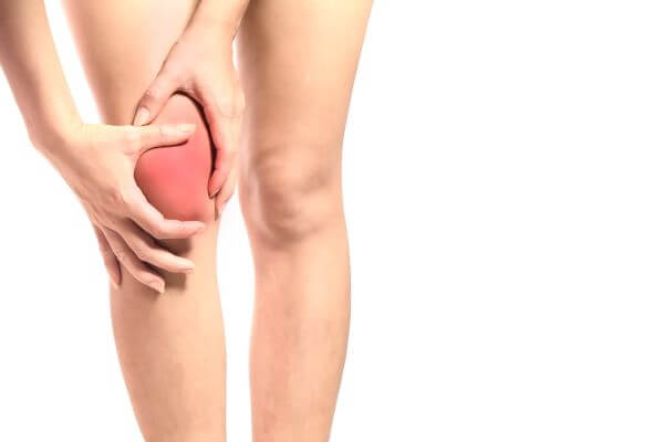
Ligament rupture produces varying degrees of pain and discomfort, knee instability and inability to bear weight on the leg. If the ligaments of the wrist or elbow joint are injured, movements such as grasping or holding objects are performed with difficulty.Ligament rupture can occur after a variety of physical activities, from dancing to snowboarding, and certain common symptoms can help identify a torn ligament as the cause of pain. At the knee, a hematoma may form along with swelling. Pain intensifies with walking or during other physical activity.
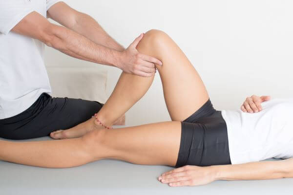
To correctly diagnose a ligament rupture, an MRI is needed to confirm the location and severity of the injury.
Types of ligament rupture
Common types of ligament injuries and tears:- Anterior cruciate ligament rupture. Of all the types of injuries seen at the knee, ACL rupture is the most common. The ACL acts as a stabilizer of the knee, preventing the tibia from sliding forward, so a tear of the ligament causes instability.
- Ankle sprain. A sprain is one of the most common conditions and can be caused by simply walking on an uneven surface. According to the American Orthopaedic Society, nearly 25,000 people sprain an ankle every day. A sprain occurs when one or more ligaments of the ankle are stretched or even torn. Symptoms include swelling of the ankle, pain and inability to bear weight. Most sprains will heal with physical rest and local ice application, but in severe cases surgery may be necessary.
- Plantar fasciitis. This is inflammation of the plantar fascia - a band of connective tissue stretched along the sole of the foot. Repetitive stress can cause micro-tears, which can lead to pain, usually located at the heel or arch of the foot. Plantar fasciitis is common among runners, and contributing factors include intense physical activity without proper warm-up and obesity.
- Acromioclavicular disjunction. This type of injury occurs following a blow or fall onto the shoulder and is a common injury of the acromioclavicular joint. It consists of partial or complete rupture of the acromioclavicular ligament.
- Grade I - mild ligament tear
- Grade II - moderate ligament tear
- Grade III - complete ligament tear
Complete ligament rupture
Complete ligament rupture has the following signs and symptoms:- pain
- bleeding/hematoma
- swelling
- joint instability
- inability to bear weight on the leg
- absence of pain (due to nerve damage, pain is not transmitted)
This type of tear generally disrupts the function and stability of the joint. Surgical treatment is recommended for patients with a complete ligament rupture. Surgical options may vary depending on the type of injury.
Partial ligament rupture
Partial ligament rupture has the following signs and symptoms:- Mild to severe pain
- Bleeding is always present
- Mild to severe swelling
- Pain with movement
- Inability to bear weight on the leg
Diagnostic methods for ligament rupture
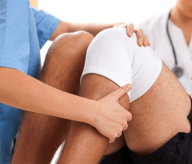
During the clinical exam, the doctor will examine the patient and the area where they report discomfort. The doctor will perform specific maneuvers to assess the general function of the joint. Often, the diagnosis can be made based solely on the clinical exam, but additional investigations may be needed to rule out other causes and determine the severity of the injury. These investigations may include:- X-ray. X-rays may be needed to rule out a bone fracture. However, X-rays cannot visualize soft tissues, such as ligaments and tendons.
- Magnetic resonance imaging (MRI). An MRI can indicate the degree of severity and signs of injury to other tissues.
- Ultrasound. Using sound waves to visualize internal structures, ultrasound can be used to examine injuries to ligaments, tendons and muscles.
Clinical examination
A clinical exam is essential when evaluating any joint. Immediately after an injury occurs, the clinical examination may be limited due to the patient's apprehension. Most injuries can be diagnosed through a detailed patient history, together with a good clinical examination. Ligament laxity can be difficult to detect in the acute phase. The Lachman test is the most sensitive test for an acute ACL tear. Since the Lachman test should be performed when the patient is relaxed, it is often better to do this test before manipulating the painful knee.While clinical examination is essential, diagnosis is often based on subjective factors, such as the physician's experience, muscle relaxation, etc. Clinical diagnosis is particularly difficult in obese patients and in patients with acute injuries and soft tissue swelling.
Imaging tests
MRI and ultrasound are now widely used to evaluate and diagnose ligament and tendon conditions. TELOS is a device that has been used to perform radiographic assessment on patients suffering from ACL injuries by analyzing the position of the tibia relative to the femur while applying a single force to the tibia. The objective results provided by this type of investigation are interesting, but the radiological technology required makes it very invasive for the patient.Injection with a special dye can also help detect damaged ligaments. This type of investigation is called an arthrogram.
Arthroscopy can be used to diagnose ligament rupture, but, being a surgical procedure, it is an invasive investigation. It is performed using an arthroscope that is inserted into the joint through a small incision, so that the ligament can be visualized directly on a computer screen.
Complications of ligament rupture
Ligament injury can range from mild (stretched ligament), moderate (partially torn), to severe (completely torn). When inadequate treatment is applied or activity is resumed prematurely, complications such as chronic pain, persistent inflammation, stiffness, instability and nerve dysfunction can occur.Chronic pain is the most common complication. Even in cases of severe injury, if pain persists and is accompanied by inflammation, there is a likelihood of an undiagnosed fracture, cartilage injury or tendon injury. Chronic pain without inflammation occurs in the case of nerve damage. Chronic pain frequently disrupts walking, which can affect the hip, knee and spine.
Persistent inflammation is caused by several conditions, but the most common cause is an untreated ligament rupture. Chronic inflammation is sometimes caused by synovitis - inflammation of the synovial membrane lining the joint capsule. Persistent inflammation in the joints is usually accompanied by low-grade pain, but it can sometimes be painless.
Nerve injuries can occur either after trauma to the knee or ankle, or as a result of chronic inflammation, which leads to increased pressure on the nerves surrounding the area. Symptoms include burning-type pain, paresthesia and muscle hypotonia, all of which are usually worsened by walking. Muscle hypotonia often causes injuries due to reduced coordination. Proprioception can also be affected, leading to imbalance and instability of the foot.
First aid in case of ligament rupture
The main cause of ligament rupture is a direct blow to the joint or an accidental fall. Timely first aid measures, aimed at reducing pain and inflammation, can significantly contribute to a faster recovery.The ankle joint is the most affected, followed by the knee and the shoulder. Knee ligament injuries are, without a doubt, the most severe, painful and debilitating. In addition, severe injuries can result in a complete ligament rupture, in which case the patient will no longer be able to move the affected area without assistance.
Cold local applications, immobilization, elevation and physical rest are the common recommendations for ligament injury. The affected area is kept in an elevated position. To reduce inflammation, ice is applied locally, which should be removed after 10-15 minutes and reapplied after 5 minutes. Anti-inflammatory medications can also help reduce pain. Immobilization is very important because any movement in the affected area can worsen the injury.
Most minor injuries will resolve after a few days of physical rest. Severe injuries, however, must be monitored by a doctor. It is almost impossible to determine the degree of severe injury by palpation or sight, and overlooking a major problem can lead to a lifelong disability. The clinical examination provides little information about the severity of the injury and there is a risk of incorrect diagnosis. In some cases, surgery is necessary to reconstruct the ligament.
In case of an accident, it is important to immobilize the affected area and transport the patient to a medical facility.
How ligament rupture is treated
Treatment for ligament rupture begins with physical rest, locally applied ice, immobilization and elevation. The patient is asked to avoid physical activities for at least two days after the injury, to prevent further damage to the ligament. If the knee or ankle is involved, the patient is advised not to bear weight on the leg for at least four days after the injury.The next step is applying ice wrapped in a towel to the affected area for 15-20 minutes, two to three times a day, to ease pain and inflammation and allow the injury to heal. During this time, the patient is advised to keep the affected area elevated to prevent swelling. These recommendations should be followed during the first three days after the accident.
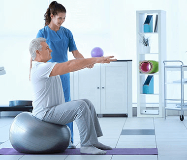
A patient suffering from a ligament rupture may be given pain relievers to control the pain. After a few days of physical rest, once the inflammation has decreased, physical rehabilitation can begin to facilitate a quick return to normal activities. The physiotherapist will design a detailed rehabilitation plan, which will include various stretching and strengthening exercises, together with a range of motion exercises to regain joint function.
Most cases of ligament rupture require a recovery time of about 4 weeks, after which the patient can return to a normal level of activity, but in some more severe cases it can take up to 12-16 weeks.
Treatment for ligament rupture depends on the severity and location of the injury. In young, athletic people, a cruciate ligament rupture is usually operated on.
If possible, conservative treatment is recommended first. If this does not bring benefits, surgery will most likely be necessary. Physiotherapy exercises are started early to train the muscle tissue and improve proprioception/coordination.
Even though pain after the injury is severe at first, symptoms are almost always eased after a few days of physical rest. If blood vessels have been damaged, a hematoma may form, which usually causes persistent pain.
Treating partial ligament rupture
In the initial recovery phase, it is recommended that the patient not bear weight on the affected leg. Depending on the ligament injuries, braces are used to help reduce pain and avoid stretching the ligament.Physiotherapy is recommended for all partial ligament ruptures, to fully restore the function of the limb and prevent possible instability that predisposes to further injuries.
Physical rest is important. However, the patient must also begin performing light exercises with the affected limb, so that the joint does not become stiff and unstable over time. Working with a physiotherapist, the patient will perform certain exercises that will increase the flexibility and strength of the affected joint.
Physical therapy and physiotherapy
Physiotherapy is a treatment that helps improve mobility (such as walking, climbing stairs), relieve pain and restore motor function and general condition.
The therapist will use a combination of techniques to reduce pain and stimulate your coordination, strength and flexibility. You may need to use exercise equipment, such as a stationary bike and treadmill.
The therapist may also treat the injury with heat or ice. Other methods that may be used include electrical stimulation, ultrasound or massage.
Exercises and stretches are done as part of a home movement program. Together with the rehabilitation program done with the therapist, home exercises speed up and enhance healing.
Wearing braces
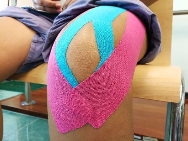
A brace can be made from different materials, elastic components or even alloys. A brace can be used in combination with, or instead of, a support band.Using a brace can be very important in preventing accidents, minimizing abnormal movements of the knee or ankle and providing the support you need. An ideal brace controls flexion and extension, while reducing harmful movements, but the best brace must meet the specific needs of the patient.
A mobile knee brace can help both prevent and treat ligament rupture. Due to its versatility, the brace is dual-purpose and can be used either as a functional band or as a prophylactic knee band. More specifically, it can help in the case of a rupture or reconstruction of the following ligaments:
- Anterior cruciate ligament (ACL)
- Lateral collateral ligament (LCL)
- Medial collateral ligament (MCL)
- Posterior cruciate ligament (PCL)
- Combined injuries (CI)
Complementary treatments
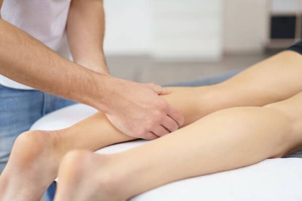
Massage therapy techniques have the ability to reduce inflammation, improve venous circulation and joint mobility, as well as ease pain. Therapeutic massage is generally considered safe by the medical community and is recognized as an effective complementary treatment for many chronic muscle and joint problems.Acupuncture was first used in ancient China, as far back as 960 AD. The word acupuncture derives from a Latin word (acus) meaning "needle" and describes puncturing soft tissues with special needles to relieve pain. According to acupuncture theory, the flow of energy (chi) circulates in the body along twelve major pathways, called meridians, each of which is connected to specific internal organs. By inserting very fine, sterile, single-use needles at certain points along the meridians, the acupuncturist can redirect and reposition the flow of energy in order to relieve tension, stress and pain. The uninterrupted and balanced flow of energy along these meridians contributes to a person's overall health.
Prolotherapy works like certain types of acupuncture, applying injections of medicinal plants and dextrose or sugar, to produce microscopic injuries to the ligaments and stimulate a new healing cycle. Electrotherapy and local modalities are used to help reduce pain and stimulate the natural healing response through electrical, magnetic, sound, light, and temperature energy.
Electrotherapy and local modalities have short-term benefits, but help when combined with other therapy methods, such as physiotherapy.
Surgical treatment for complete ligament rupture
Unfortunately, when the ligament is completely torn, surgery is recommended, as it will not regenerate on its own.Currently, many techniques are used by orthopedic surgeons around the world to perform this operation, but the principle behind each remains relatively the same.
In the case of a completely torn anterior cruciate ligament, the newest surgical technique uses the arthrometer. An arthrometer is an instrument designed for the objective evaluation of the ACL using a reproducible and non-invasive method. Currently, orthopedic surgeons choose to work with the arthrometer to optimize the treatment they provide to patients suffering from an ACL injury. These advanced devices provide valuable information about the state of the ACL before choosing surgery and allow precise monitoring of the ACL graft after the operation.
Ligament injuries that are recognized early enough (within a few weeks of the injury) can be repaired by inserting metal pins to stabilize the bones, which allows the ligaments to heal. Once healed, the pins are removed. This is less likely to be useful if too much time has passed since the injury.
Arthroscopic surgery can be used to access and repair ligaments. Arthroscopy allows the surgeon to see the anatomy of the joint and the movement of the ligament without the need for large incisions.
The right time for surgery
The doctor may recommend surgery if:- The patient is an athlete and wants to continue playing sports, especially if the sport involves jumping or pivoting
- More than one ligament or cartilage in the knee is injured
- The patient is young and active
- The injury causes knee instability during daily activities
Surgery by suture or graft
For injuries that occurred six months or more before treatment, ligament reconstruction may be necessary. A tendon graft is used to replace the torn ligament, and the injured area is temporarily held in position with metal pins throughout the initial healing process.During reconstruction, the surgeon removes the damaged ligament and replaces it with a segment of tendon, tissue similar to that of the ligament, called a graft.
Postoperative recovery
After surgery, patients wear a splint or brace at first. Once these are removed, some patients notice stiffness in the affected area, while others notice a limited range of motion. Manual therapy is often incorporated as part of the recovery process to ensure proper joint alignment and function recovery during healing. Athletes can often return to sports after 8 to 12 months.The major goals of rehabilitation include:
- Achieving good functional stability
- Regaining muscle strength
- Achieving the best possible functional level
- Reducing the risk of recurrence
Closed kinetic chain exercises have become more popular than open kinetic chain exercises in ACL rehabilitation. Therapists consider CKC exercises to be safer than OKC exercises, since they place less stress on the graft. In addition, CKC exercises are considered to be more functional and just as effective as OKC exercises.
Measures for preventing ligament rupture
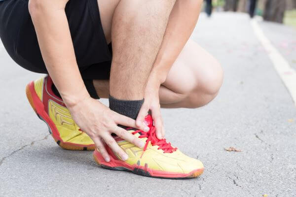
Successful ligament rupture prevention programs may differ in their exercises, but share a common focus: improving flexibility, strength (especially of the abdomen, hips and legs), balance, agility and the ability to jump and land safely.Develop strength in the hips and thighs. Warm up and stretch before sports and training. Increase blood flow in the muscles before starting an exercise program. Being flexible enough to move freely can help you maintain proper form. Include stretches for the thighs, calves and hips, and pay special attention to any areas that are particularly tight.
Physical exercises to reduce the risk of ligament rupture
Weight control and the risk of ligament rupture
Measures for preventing ligament rupture in high-risk sports
The best way to prevent an ACL injury is to strengthen the muscles around the knee, especially the hamstrings and quadriceps. These two muscle groups are crucial for regaining balance and control. Strengthening the hamstrings and quadriceps can help prevent knee twisting and hyperextension, which can contribute to ligament rupture.
Strength training is an important way to help prevent injuries. Others include:
- maintaining control when practicing sports such as skiing and basketball.
- bending the hips and knees to cushion the landing after a jump.
- warming up before doing sports.
Avoiding high-risk movements for ligament rupture
Common causes of spinal ligament rupture are weightlifting, sports contact, racket sports that require twisting, improper posture that positions the spine out of alignment, and pregnancy and weight gain, both of which can put excessive pressure on the spinal ligaments. This type of injury can be prevented by doing strengthening and stretching exercises. Strengthening the abdominal muscles also helps with proper spinal posture and relieves pressure on the ligaments.
High-heeled shoes and the risk of ligament rupture
When you wear high heels, the feet are in a permanent plantar flexion, so the tibiofibular ligaments are under continuous stretch. Wearing high heels affects the tibiofibular ligaments, the metatarsals, the knees and the back. To avoid the negative effects, you should not wear heels higher than 4 cm, and when you do wear high heels, try not to stand in them for more than 2 hours.
IF YOU DON'T ALREADY KNOW US
Centrokinetic is the leading medical clinic dedicated to bone and joint conditions, organized into several dedicated departments:
- Orthopedics: made up of a highly experienced team of orthopedic surgeons, led by Dr. Andrei Ioan Bogdan, senior orthopedics-traumatology physician with surgical activity at the Floreasca Clinical Emergency Hospital and at the Medlife Orthopedic Hospital, specialized in sports traumatology and in ankle and foot surgery.
- Pediatric orthopedics, where we treat children's sports injuries (ligament and meniscus injuries), spinal deformities (scoliosis, kyphosis, hyperlordosis), and foot conditions (hallux valgus, hallux rigidus, clubfoot, flat valgus foot, cavus foot).
- Neurology, providing consultations and EMG within the department. The medical team is completed by Dr. Mardare, highly specialized at the University of Oxford in spinal surgery.
- Medical rehabilitation, for children and adults, the department being led by one of the most experienced physicians in the field, senior lecturer Dr. Tache Georgiana. The department specializes in the recovery of performance athletes, in spinal conditions, and in the recovery of children with neurological and traumatic conditions.
- Medical imaging, the clinic being equipped with ultrasound and MRI, dedicated to musculoskeletal conditions. We are proud of our medical team of radiologists, made up of Dr. Ghiea Sorin, Dr. Pantu Cosmin, Dr. Florescu Nicoleta, specialized in musculoskeletal imaging.
Find out the latest news about us by following the Facebook and YouTube accounts of the Centrokinetic clinic.

MAKE AN APPOINTMENT
CONTACT US
MAKE AN APPOINTMENT
FOR AN EXAMINATION
See here how you can make an appointment and the location of our clinics.
MAKE AN APPOINTMENT
BUCHAREST TEAM


































































CLUJ NAPOCA TEAM


































BRASOV TEAM


















Asiguratori privati





Parteneriate



CENTROKINETIC FLOREASCA
Bucharest, 1st District,
Mircea Eliade Street, no. 18,
Eliade Tower, Entrance A, Floor 3

| Navigation | Explore 3D |
CENTROKINETIC Barbu Vacarescu
Bucharest, 2nd District,
Barbu Vacarescu Street, no. 102,
102 The Address, ground floor.

| Navigation | Explore 3D |
CENTROKINETIC MILITARI
Bucuresti, Sector 6,
Bld. Iuliu Maniu, nr. 15G,
Cladirea PBT, etaj 3.

| Navigation |



CENTROKINETIC CLUJ-NAPOCA
Cluj-Napoca,
Str. Intre Lacuri, nr.1
Intrarea este in spatele cladirii.

| Navigation |


SOCRATES CLINIC TIMISOARA
Timisoara, Calea Martirilor 1989,
nr. 1.
Telefon: 0371 785 374.

| Navigation |
CONTACT
Phone: 0319693
E-mail: programari@centrokinetic.ro
Operating hours:
Monday – Friday: 07:00 – 21:00
Saturday: 09:00 – 14:00
Sunday: closed.
MAKE AN APPOINTMENT

