
MUSCULOSKELETAL ULTRASOUND


- Musculoskeletal ultrasound – general information
- What is musculoskeletal ultrasound
- What an ultrasound machine is and how it works
- When musculoskeletal ultrasound is recommended
- Body areas that can be examined with musculoskeletal ultrasound
- Conditions that can be detected with a musculoskeletal ultrasound
- Contraindications for performing musculoskeletal ultrasound
- Advantages of musculoskeletal ultrasound
- Limitations of musculoskeletal ultrasound
- Performing the musculoskeletal ultrasound
- Complementary tests to musculoskeletal ultrasound
Musculoskeletal ultrasound – general information
Musculoskeletal ultrasound changes the treatment and recovery of sports injuries and other muscle, tendon and ligament problems. If you suffer from pain in any of your joints, then ultrasound will help you get an accurate diagnosis and evaluate effective treatments, such as surgery, monitoring therapy and assisted physiotherapy treatments.This is a safe, noninvasive technique that is fast and precise in diagnosing many problems. Depending on the clinical symptoms presented, the ultrasound exam may involve a full evaluation of the joint or may be tailored to a specific region of interest.
Centrokinetic provides musculoskeletal ultrasound services, with the role of diagnosing many conditions, from ligament tears to disc herniations, as well as precisely guiding the needle during injections.
What is musculoskeletal ultrasound
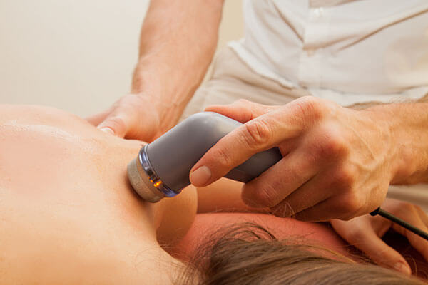
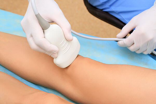
Ultrasound of the musculoskeletal system provides images of the muscles, tendons, ligaments, joints and soft tissues throughout the body. This type of scan is commonly used to help diagnose tendon tears, such as rotator cuff tears in the shoulder or Achilles tendon tears in the ankle, muscle abnormalities such as tears and soft tissue masses, bleeding or fluid collection inside muscles, or damage to major joints. It can also be used as a guide for steroid injections into joints and surrounding soft tissue, as an effective treatment for painful joints. No special preparation is needed for these types of scans.Musculoskeletal ultrasound for clinical diagnosis is painless, safe and does not involve radiation. It simply uses sound waves to produce clear images of the inside of the body. The procedure involves an ultrasound probe, called a transducer, which looks like a microphone. A trained physician applies gel to the skin and moves the instrument over the affected area. The ultrasound transducer emits and receives high-frequency sound waves, which cannot be heard by the human ear. The sounds that return are recorded, and a computer then uses these sounds to create an image. This powerful diagnostic tool can provide valuable information for a physiotherapist.
What an ultrasound machine is and how it works
A basic ultrasound machine has the following components:- Transducer probe - the probe that sends and receives sound waves
- Central processing unit (CPU) - the computer that does all the calculations and contains the power supplies
- Transducer control pulse - modifies the amplitude, frequency and duration of the pulses emitted by the transducer probe
- Display - displays the image from the ultrasound data processed by the CPU
- Keyboard / cursor - enters data and measures the display
- Disk storage device (hard drive, floppy, CD) - stores the obtained images
- Printer - prints the image from the displayed data
Transducer probes come in several shapes and sizes. The shape of the probe determines its field of view, and the frequency of the emitted sound waves determines how deeply the sound waves penetrate and what the image resolution is. Transducer probes may contain one or more crystal elements; in multi-element probes, each crystal has its own circuit. Multi-element probes have the advantage that the ultrasound beam can be "steered" by changing the timing at which each element is pulsed; beam direction is especially important for cardiac ultrasound. In addition to probes that can be moved over the surface of the body, some probes are designed to be inserted through various body openings so they can get closer to the organ being examined (uterus, prostate gland, stomach); getting closer to the organ can allow more detailed views.
When musculoskeletal ultrasound is recommended
There are several advantages to musculoskeletal ultrasound compared to other types of imaging, but usually the doctor will order several tests that complement each other. For example, for an injury you can have both an MRI exam and a musculoskeletal ultrasound.However, ultrasound offers a unique analysis of an injury. It allows the technician to easily capture an image from several views. This is especially useful if the symptoms you are experiencing are triggered or worsened by movement. For example, maybe the shoulder is not painful if it is not moved beforehand. With a musculoskeletal ultrasound, the doctor can see what is happening with the joint at that moment. With other forms of imaging, you must remain still for the image to be clear, but ultrasound can capture this movement.
Through ultrasound technology, the doctor can visualize more complex structures in the body to detect abnormalities as well. Imaging tests such as an MRI may miss tiny details.
A musculoskeletal ultrasound also offers patients some benefits. Imaging is done in an open space, so it is a practical choice for patients who cannot tolerate a traditional MRI.
Musculoskeletal ultrasound is usually ordered to diagnose:
- Tears in ligaments, muscles and tendons
- Bleeding in muscles or joints
- Fluid accumulation
- Soft tissue tumors
Musculoskeletal ultrasound is a noninvasive test, with no risk to the patient, which gives the doctor one of the best ways to visualize the problem and create a solution for it.
Body areas that can be examined with musculoskeletal ultrasound
Common areas that can be examined with musculoskeletal ultrasound include the shoulder, elbow, wrist and hand, hip, knee and ankle. It can be especially useful following a sports accident.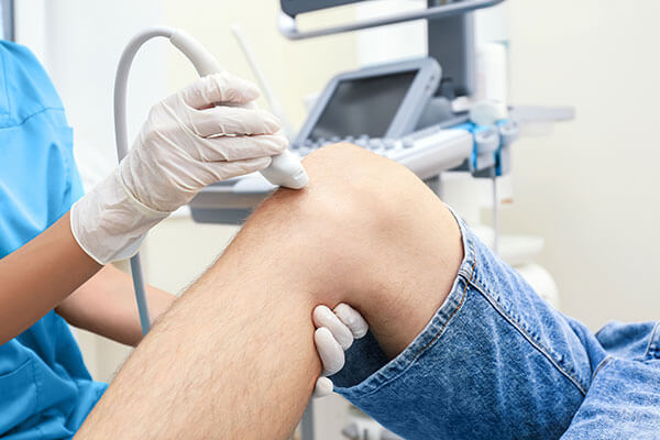
Conditions that can be detected with a musculoskeletal ultrasound
Musculoskeletal ultrasound is used in orthopedic practice for diagnosing injuries, as well as for guiding the needle during injections. Musculoskeletal ultrasound is frequently used to diagnose:- Tendon tears or tendinitis.
- Muscle disorders, masses or fluid collections.
- Ligament tears or sprains.
- Nerve entrapment.
- Inflammation or fluid in the bursa and joints.
- Early changes of rheumatoid arthritis.
- Hernia.
- Benign and malignant soft tissue tumors.
- Ganglion cysts.
- Foreign bodies in soft tissue (such as splinters or glass).
- Hip dislocations in infants.
- Shoulder and back - rotator cuff tendon tears or tendinitis in the shoulder, frozen shoulder, pain, swelling or masses, impingement and instability.
- Elbow - ligament tears or sprains, golfer's or tennis elbow, muscle tears, masses or fluid collections.
- Wrist and hand - pain, swelling or inflammation of the joints, soft tissue injuries, nerve entrapment in carpal tunnel syndrome, masses, rheumatoid arthritis.
- Groin region - pain, swelling, inguinal / femoral hernia.
- Lower back - pain, osteoarthritis.
- Knee - pain, cruciate cartilage injuries, joint swelling, injuries, masses.
- Ankle and foot - Achilles tendon disorders, injuries, instability, impingement, numbness, tingling, masses or swelling.
Contraindications for performing musculoskeletal ultrasound
Musculoskeletal ultrasound has no contraindications, is dynamic, safe and has few technical limitations.There are no known harmful effects for standard musculoskeletal ultrasound. In any case, ultrasound has difficulty penetrating bone and, therefore, can see only the outer surface of bone structures and not what is inside. For visualizing the internal structure of bones or certain joints, other imaging modalities such as MRI are usually used.
Advantages of musculoskeletal ultrasound
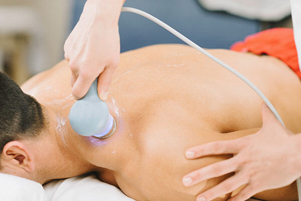
There are major advantages to using musculoskeletal ultrasound over other imaging modalities:Every patient can undergo ultrasound - There are no contraindications for ultrasound. Even patients with metal implants or cardiac implants can undergo the examination (which is not possible with MRI). Ultrasound examinations are performed with the patient in a more comfortable position.
Allows real-time dynamic examination of the patient - Static imaging may not show tissues and structures in motion, when pain and dysfunction can occur. During an ultrasound examination, the patient can perform dynamic movements that cause pain, and doctors can observe in real time how the structures respond. Dynamic examinations can show changes in conditions such as shoulder impingement that would not appear on a static MRI.
Facilitates rapid bilateral comparison - Doctors performing ultrasound examinations are able to quickly evaluate contralateral limbs to detect bilateral asymmetries and to assess whether pathology exists or whether it is simply reduced to anatomical variability.
Most ultrasound scans are noninvasive (no needles or injections) and are usually painless. Ultrasound is widely available, easy to use and less costly than other imaging methods. Ultrasound imaging does not use any ionizing radiation.
Limitations of musculoskeletal ultrasound
Musculoskeletal ultrasound is limited by the fact that it does not penetrate bone to any usable extent and, therefore, can only show the outer surface of bone structures. In infants, however, the extensive presence of cartilage still allows musculoskeletal scanning of bone surfaces. The cartilage surrounding the hip and shoulder muscle is also difficult to scan due to its proximity to the bone. Bone tumors and other solid masses must be evaluated using other methods, and soft tissue tumors are better evaluated using MRI. Moreover, ultrasound is not useful in diagnosing certain types of back pain.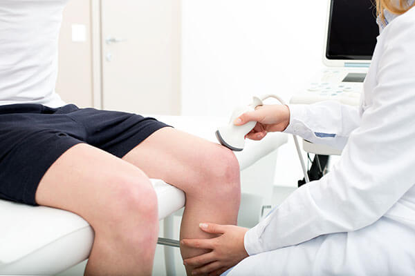
How musculoskeletal ultrasound is performed
During a musculoskeletal ultrasound, warm gel is placed on the area to be tested and a probe is gently pushed back and forth over the patient's skin to create and pick up sound waves, which are converted into an image on the screen. The gel helps the transducer or scanning probe ensure secure contact with the body and allow ease of movement of this probe over the examined area. Images will be captured from any problem area. Typical scans take 15-30 minutes. If the patient is having an injection, there may be additional time for preparation, setup, observation after the injection, etc. All procedures are performed in the office.During the scan you will discuss your clinical symptoms and will be asked to demonstrate or describe the mechanisms of injury, point to areas of tenderness, and show whether there are symptomatic changes with movement.
Depending on the area being examined, you will be seated or lying on a bed or on a swivel chair. You may be asked to move the examined extremity to evaluate the anatomy and function of the joint, muscle, ligament or tendon.
When imaging is complete, the clear gel will be wiped off the skin. If left on the skin, it will dry into a powder and will not stain or discolor clothing. Once the exam is complete, you can resume your normal level of activity.
Preparing the patient for a musculoskeletal ultrasound
Fortunately, this will be a quick, simple and painless process. No preparation is necessary, but you will need to change into a gown for the procedure. The tips below will help you:- Wear clothing and footwear that is easy to take off.
- Leave jewelry at home, if possible.
- Plan to be there for about an hour. While the ultrasound procedure itself is quite quick, you may be asked to stay while the images are reviewed.
Ultrasound procedure
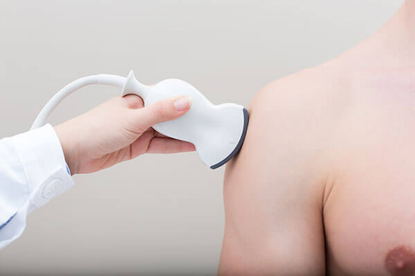
At the examination, you will need to remove all clothing and jewelry from the area to be examined and you may wear a gown during the procedure. You will usually lie on your back on an examination table, but for special areas you may be placed in different positions. The probe will be moved back and forth over the relevant part of the body to scan the area of interest. You may also be asked to move your joints or limbs during the scan, so the sonographer can observe the affected area while it is in motion.Although the exam is safe and painless, the patient must remain still since the exam is sensitive to movement. Apart from what was mentioned, no other preparation is required from the patient.
Interpreting the results
Musculoskeletal ultrasound is performed by assistant physicians and doctors. The results are interpreted by the examiner at the time of the visit. The results are discussed and a treatment plan is made.Doctors are trained to administer therapeutic injections under ultrasound guidance, improving effectiveness, leading to decreased pain and better results.
Complementary tests to musculoskeletal ultrasound
Ultrasound may find that further examinations or other imaging techniques may be necessary to further evaluate a problem area. Sometimes an X-ray, MRI or CT scan may be needed.Musculoskeletal Ultrasound Prices
You can find here a detailed list of the prices for individual services. However, any proper recovery process is based on a mixed plan of therapies and procedures, personalized according to the condition, stage, patient profile, and other objective medical factors. As a result, to set up a treatment plan, with the therapies involved, the periods and the corresponding rate for the plan, please book an appointment here for an initial consultation.
IF YOU DON'T ALREADY KNOW US
Centrokinetic is the leading medical clinic dedicated to bone and joint conditions, organized into several dedicated departments:
- Orthopedics: made up of a highly experienced team of orthopedic surgeons, led by Dr. Andrei Ioan Bogdan, senior orthopedics-traumatology physician with surgical activity at the Floreasca Clinical Emergency Hospital and at the Medlife Orthopedic Hospital, specialized in sports traumatology and in ankle and foot surgery.
- Pediatric orthopedics, where we treat children's sports injuries (ligament and meniscus injuries), spinal deformities (scoliosis, kyphosis, hyperlordosis), and foot conditions (hallux valgus, hallux rigidus, clubfoot, flat valgus foot, cavus foot).
- Neurology, providing consultations and EMG within the department. The medical team is completed by Dr. Mardare, highly specialized at the University of Oxford in spinal surgery.
- Medical rehabilitation, for children and adults, the department being led by one of the most experienced physicians in the field, senior lecturer Dr. Tache Georgiana. The department specializes in the recovery of performance athletes, in spinal conditions, and in the recovery of children with neurological and traumatic conditions.
- Medical imaging, the clinic being equipped with ultrasound and MRI, dedicated to musculoskeletal conditions. We are proud of our medical team of radiologists, made up of Dr. Ghiea Sorin, Dr. Pantu Cosmin, Dr. Florescu Nicoleta, specialized in musculoskeletal imaging.
Find out the latest news about us by following the Facebook and YouTube accounts of the Centrokinetic clinic.

MAKE AN APPOINTMENT
CONTACT US
MAKE AN APPOINTMENT
FOR AN EXAMINATION
See here how you can make an appointment and the location of our clinics.
MAKE AN APPOINTMENT


BUCHAREST TEAM


































































CLUJ NAPOCA TEAM


































BRASOV TEAM
























Asiguratori privati





Parteneriate



CENTROKINETIC FLOREASCA
Bucharest, 1st District,
Mircea Eliade Street, no. 18,
Eliade Tower, Entrance A, Floor 3

| Navigation | Explore 3D |
CENTROKINETIC Barbu Vacarescu
Bucharest, 2nd District,
Barbu Vacarescu Street, no. 102,
102 The Address, ground floor.

| Navigation | Explore 3D |
CENTROKINETIC MILITARI
Bucuresti, Sector 6,
Bld. Iuliu Maniu, nr. 15G,
Cladirea PBT, etaj 3.

| Navigation |



CENTROKINETIC CLUJ-NAPOCA
Cluj-Napoca,
Str. Intre Lacuri, nr.1
Intrarea este in spatele cladirii.

| Navigation |


SOCRATES CLINIC TIMISOARA
Timisoara, Calea Martirilor 1989,
nr. 1.
Telefon: 0371 785 374.

| Navigation |
CONTACT
Phone: 0319693
E-mail: programari@centrokinetic.ro
Operating hours:
Monday – Friday: 07:00 – 21:00
Saturday: 09:00 – 14:00
Sunday: closed.
MAKE AN APPOINTMENT

