
Leziunea Bankart


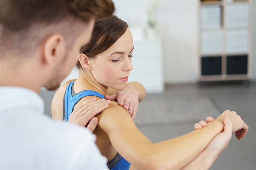
- General information about the Bankart lesion
- Shoulder anatomy
- When the Bankart lesion occurs
- Causes
- Symptoms
- Feeling of shoulder instability
- Repeated dislocations
- Shoulder pain
- Diagnosis
- Clinical examination
- Imaging tests
- Treatment
- Postoperative medical recommendations
- Recovery
- Chances of a repeated dislocation
This article was written by Dr. Catalin Vlad Gican, orthopedic specialist, with over 10 years of experience, highly specialized in sports traumatology.
General information about the Bankart lesion
The labrum of the joint, the second type of cartilage in the shoulder, can be torn as a consequence of a strong shoulder dislocation. When the damage involves only the labrum, it is called a Bankart lesion. A Bankart lesion is an injury to the anterior part of the glenoid labrum of the shoulder. This injury is caused by repeated subluxations of the anterior shoulder. Dislocation of the shoulder joint (anterior) can damage the ring of connective tissue around the glenoid labrum. It can also affect the connection between the labrum and the capsule. It usually has to do with a weak construction of the medial glenohumeral ligament. This injury is common among athletes who play volleyball, tennis, handball and people who perform overhead arm-raising activities.Shoulder anatomy
The shoulder is one of the largest and most complex joints in the body. The shoulder joint is formed where the humerus (the upper arm bone) fits into the scapula (the shoulder blade). Other important bones in the shoulder include:• The acromion is a bony projection from the scapula.
• The clavicle meets the acromion at the acromioclavicular joint.
• The coracoid process is a hook-like bony projection from the scapula.
The shoulder has several important structures:
• The rotator cuff is a collection of muscles and tendons that surround the shoulder, providing support and allowing a wide range of movement.
• The bursa is a small fluid-filled sac that cushions and protects the rotator cuff tendons.
• The cartilage rim called the labrum forms a cup for the humeral head to fit into.
• The humerus fits relatively loosely in the shoulder joint. This gives the shoulder a wide range of motion, but also makes it vulnerable to injury.
When the Bankart lesion occurs
Bankart lesions occur as a direct result of anterior dislocation of the humeral head, during which the humerus is compressed against the labrum. There is detachment of the anterior-inferior labrum from the inferior glenoid, and the labral tear can extend further up or back. "Soft" Bankart-type lesions are more common than "bony" Bankart lesions.Causes
The shoulder is designed for its mobility, with stability being sacrificed to achieve this mobility. Due to the poor bone fit and capsular laxity, the glenohumeral joint is highly unstable, making it the most frequently dislocated joint in the human body. It relies on dynamic stabilizers and the neuromuscular system for its stability. Anterior instability is the most common type of traumatic instability, accounting for about 95% of all shoulder instabilities. Glenohumeral dislocations are mainly caused by an extension and external rotation movement. In many cases of anterior dislocation, patients have a Bankart lesion. A reverse Bankart lesion can occur in the case of a posterior dislocation.The same mechanisms responsible for shoulder dislocation are among the causes leading to Bankart lesions. These pathologies are most often diagnosed together with an anterior shoulder dislocation, and in patients with recurrent shoulder dislocations. Bankart lesions are common in young men involved in contact sports, and in women over 60 years old due to falls.
Any physical activity or sport that predisposes the body to a shoulder dislocation presents a risk for Bankart lesions. These include people who actively play contact sports (soccer, rugby, hockey) and throwing activities (tennis, baseball, cricket). The increased incidence of falls onto the shoulder presents a higher risk in older people. The poor vascularization of the glenoid labrum exposes this area to injury when it is strained. These pathologies can easily occur in patients with common congenital laxity, a condition called Ehlers-Danlos Syndrome, which weakens the ligaments that stabilize the joints.
Symptoms
The most common symptom of Bankart lesions is a feeling of instability of the shoulder joint, especially if these pathologies occur after repeated dislocations. The patient may display excessive displacement of the humeral head in the glenoid joint. Pain and a sensation of catching in shoulder subluxation are also frequently described. Patients often state that when they move their arm behind their head, they feel that the shoulder is almost dislocated.For a correct diagnosis of this injury, schedule a consultation at Centrokinetic.
Feeling of shoulder instability
The most common type of shoulder instability is traumatic anterior instability. In this type of instability, the ligaments and labrum at the front-lower part of the shoulder are torn by an injury that occurred when the arm was stretched to the side. The labrum and ligaments often do not heal completely, and the shoulder continues to feel unstable (for example, when the arm is moved to the side and back). These injuries seriously compromise shoulder stability. An unhealed Bankart lesion can lead to recurrent anterior shoulder instability. When multiple dislocations occur, the chances of healing without surgery become small.A similar type of injury can occur at the back of the joint (traumatic posterior shoulder instability), but it is much less common. Traumatic posterior instability results from mechanisms such as a fall onto an outstretched hand.
There is another type of instability that occurs without an injury - atraumatic instability. In this condition, the shoulder loses its normal ability to center the ball in the glenoid socket. Not infrequently, atraumatic instability can allow the shoulder to slide in different directions (multidirectional instability). In this condition there is usually nothing torn, but rather the shoulder's stabilizing structures decompensate.
Repeated dislocations
A subluxation can be more difficult to identify than a complete dislocation. However, in some cases, the partially dislocated humerus is visible under the skin.A person may feel the movement of the humeral head in and out of the socket, which is usually uncomfortable and can be painful. Once a shoulder is dislocated, the joint can become unstable and prone to repeated dislocations or subluxations.
Shoulder dislocations can be partial, with the ball of the upper arm coming only partially out of the socket. This is called a subluxation. A complete dislocation means that the ball is completely out of the socket.
Once the ligaments, tendons and muscles around the shoulder become weakened or torn, dislocations can occur repeatedly. Chronic shoulder instability is the persistent inability of these tissues to keep the arm centered in the shoulder socket.
Shoulder pain
Shoulder pain due to a Bankart lesion can occur when the patient tries to raise their arm overhead, at night, or during daily activities. Also, trying to throw a ball can cause pain. Bankart-type tears usually cause pain in the front of the shoulder and can be associated with catching or locking sensations, or a feeling of the shoulder slipping. The onset of symptoms usually occurs after a traumatic event, such as a shoulder dislocation. Examination of the shoulder shows pain with compression and rotation of the shoulder, and may reveal instability when the shoulder is tested for laxity.Diagnosis
A medical history establishes the beginning of a clinical investigation to assess the possible causes, injuries and previous shoulder dislocations that led to a recurrent shoulder dislocation. A high-energy trauma event, especially in young active people, presents a higher risk for a Bankart lesion, requiring a thorough medical evaluation. The clinical exam provides a good indication of a dislocated shoulder and, possibly, other associated pathologies. Specific functional tests include the apprehension test, the relocation test and the sulcus sign. However, radiological imaging is necessary for the exact diagnosis of a Bankart lesion. A Bankart lesion of the labrum is best visualized by MRI, using a contrast agent. X-rays and CT scans do not easily detect a Bankart lesion since it involves soft tissue, but they are useful for diagnosing a Hill-Sachs lesion on the humeral head and possible associated fractures.Clinical examination
After discussing your symptoms and medical history, the doctor will examine your shoulder. Specific tests help your doctor assess instability in the shoulder. Your doctor may also perform tests for general ligament laxity.Imaging tests
Your doctor may recommend imaging tests to confirm the diagnosis, and to identify any other problems.X-rays. These images will show injuries to the bones that make up the shoulder joint.
Magnetic resonance imaging (MRI). This provides detailed images of soft tissues. It can help your doctor identify injuries to the ligaments and tendons around the shoulder joint.
A Bankart lesion of the soft tissue can be seen on arthroscopy and MR arthrography as a labral fragment attached to the anterior band of the inferior glenohumeral ligament, and as a tear in the periosteum of the scapula. A bony Bankart lesion can also be found on X-rays.
Treatment
Bankart lesions heal, and therefore early surgery is not necessary. There are two general options for treating a Bankart lesion. One option is to allow the arm to rest, with the inflammation decreasing through the use of a sling. This is usually followed by physical therapy to regain movement of the limb. The potential downside of this option is that people who dislocate their shoulder once are more likely to suffer repeated dislocations. Since the damaged labrum does not heal in a perfect position, the shoulder joint is less stable. Younger patients and those who participate in higher-risk sports (such as ice hockey) are at particularly high risk for repeated dislocations. For this reason, many of these patients are advised to have surgery, rather than non-surgical treatment.The other option is to perform surgery to repair the torn labrum. When surgery is performed, the torn ligament is reattached to the shoulder socket.
Non-surgical treatment
The first approach to conservative treatment for a Bankart lesion due to shoulder dislocation is temporary immobilization with a sling until the inflammation subsides. The patient requires treatment with analgesics and nonsteroidal anti-inflammatories in the acute phase and will subsequently benefit from intensive physical therapy. However, with non-surgical treatment there is a higher risk of recurrent dislocations. If the shoulder dislocates again, surgery is necessary.Surgical treatment
Surgery for a Bankart lesion is highly recommended for active athletes, even after a single dislocation, especially for those involved in contact sports, which increases the likelihood of a larger shoulder dislocation, and of shoulder joint injuries. Similar to SLAP lesions, surgery consists of repairing the labrum, followed by reattaching the glenohumeral ligament to the glenoid cavity, mainly through arthroscopic surgery. The success rate of surgical repairs is about 90%, and patients can return to their sports activities.Shoulder arthroscopy. Most often, minimally invasive procedures can be used to repair the labrum. Your doctor may use several small surgical instruments to locate and examine the tear, remove the damaged fragments and trim them. Your doctor will place 2-3 anchors on the bone near the tear. The sutures attached to the bone are pulled tight, reattaching the torn piece of labrum to the bone. Any torn muscle, tendon or cartilage will be fixed and any damaged tissue will be removed.
Open surgery. In some cases, orthopedic surgeons may prefer to use open surgery for this procedure. Which method your doctor chooses depends on the type and location of the injury, the condition of your tissue and how much additional damage has occurred.
Depending on the degree of injury, other recommended surgical procedures may include:
• Ligament reattachment
• Shaping or adding bone graft to provide stability to the glenoid (socket)
• Reshaping, building up or thickening the glenoid labrum cartilage
• Tightening the ligaments and the capsule to fix injuries and add stability
If you are healthy, the outlook for this type of shoulder surgery is generally good, with most patients resuming light movement and non-contact sports within 3 to 6 weeks. Most patients will need to avoid lifting and heavy-impact activities for at least three months, while benefiting from physical therapy to regain strength and range of motion in the shoulder.
Postoperative medical recommendations
After an arthroscopic Bankart repair, patients will generally be asked to keep their arm immobilized for about a month. However, physical therapy will begin on day 5 after the operation. In addition, patients will undergo physical therapy for about four months to strengthen the muscle tissue, and to improve the range of motion in the shoulder. Patients are often restricted from participating in contact sports for a period of six months after surgery, to allow the shoulder to fully heal.Recovery
Rehabilitation after a Bankart reconstruction usually takes 4-6 months, depending on the level of sport or activity. A shoulder immobilizer with a small pillow near the back is used to protect the area for the first six weeks.Physical therapy is initiated one week after surgery and involves a passive range of motion, in which the therapist moves the shoulder within a specific range. Active movement, in which the patient begins to move their shoulder without assistance, starts 6 weeks after surgery. There is a progression toward more aggressive strengthening at three to four months after surgery, and a return to some sports at four to six months after the operation. Returning to throwing usually takes ten to twelve months, and to swimming eight to ten months after surgery. It is important to recognize that returning to sport is based on several factors, including biological healing, the return of strength and endurance, subjective improvement and a gradual return to sport-specific activities. Therefore, the time frames for returning to sports activities can vary.
Chances of a repeated dislocation
Bankart tears are injuries associated with people who have had a shoulder dislocation and sustained a certain type of cartilage injury as a result. When a Bankart tear occurs inside the shoulder, the ball and socket of the joint tend to be unstable, and repeated dislocations represent a major concern. To prevent repeated dislocations, many people choose to undergo surgery to repair Bankart lesions.IF YOU DON'T ALREADY KNOW US
Centrokinetic is the leading medical clinic dedicated to bone and joint conditions, organized into several dedicated departments:
- Orthopedics: made up of a highly experienced team of orthopedic surgeons, led by Dr. Andrei Ioan Bogdan, senior orthopedics-traumatology physician with surgical activity at the Floreasca Clinical Emergency Hospital and at the Medlife Orthopedic Hospital, specialized in sports traumatology and in ankle and foot surgery.
- Pediatric orthopedics, where we treat children's sports injuries (ligament and meniscus injuries), spinal deformities (scoliosis, kyphosis, hyperlordosis), and foot conditions (hallux valgus, hallux rigidus, clubfoot, flat valgus foot, cavus foot).
- Neurology, providing consultations and EMG within the department. The medical team is completed by Dr. Mardare, highly specialized at the University of Oxford in spinal surgery.
- Medical rehabilitation, for children and adults, the department being led by one of the most experienced physicians in the field, senior lecturer Dr. Tache Georgiana. The department specializes in the recovery of performance athletes, in spinal conditions, and in the recovery of children with neurological and traumatic conditions.
- Medical imaging, the clinic being equipped with ultrasound and MRI, dedicated to musculoskeletal conditions. We are proud of our medical team of radiologists, made up of Dr. Ghiea Sorin, Dr. Pantu Cosmin, Dr. Florescu Nicoleta, specialized in musculoskeletal imaging.
Find out the latest news about us by following the Facebook and YouTube accounts of the Centrokinetic clinic.

PROGRAMEAZA-TE PENTRU O CONSULTATIE
Pentru ca orice tratament in clinica noastra este bazat pe un diagnostic si se face sub urmarire medicala, pentru evaluarea permanenta a evolutiei.PROGRAMEAZA-TE
PROGRAMEAZA-TE
LA O CONSULTATIE
Vezi aici modalitatile de programare si localizarea clinicii
SOLICITA O PROGRAMARE


MEDICI
BUCURESTI


































































MEDICI
CLUJ-NAPOCA


































MEDICI
BRASOV






















Asiguratori privati





Parteneriate



CENTROKINETIC FLOREASCA
Bucuresti, Sector 1,
Bd. Mircea Eliade, nr. 18.
Eliade Tower, intrarea A, Etaj 3

| Navigatie | Exploreaza 3D |
CENTROKINETIC Barbu Vacarescu
Bucuresti, Sector 2,
Str. Barbu Văcărescu, nr. 102
102 The Address, Parter

| Navigatie | Exploreaza 3D |
CENTROKINETIC MILITARI
Bucuresti, Sector 6,
Bld. Iuliu Maniu, nr. 15G,
Cladirea PBT, etaj 3.

| Navigatie |



CENTROKINETIC CLUJ-NAPOCA
Cluj-Napoca,
Str. Intre Lacuri, nr.1
Intrarea este in spatele cladirii.

| Navigatie |



CONTACT
Telefon: 0319693
Email: programari@centrokinetic.ro
Orarul de functionare:
Luni – Vineri, intre 07:00 – 21:00
Sambata, intre 09:00 – 14:00

