
RECONSTRUCTION OF THE SIDE EFFECTS OF THE ANKLE
For all traumatic or chronic diseases of the musculoskeletal system, the Centrokinetic private clinic in Bucharest is prepared with an integrated Orthopedic Department, which offers all the necessary services to the patient, from diagnosis to complete recovery.
The Department of Orthopedic Surgery of the Centrokinetic Clinic is dedicated to providing excellent patient care and exceptional education for young physicians in the fields of orthopedic surgery and musculoskeletal medicine.
Discover the open MRI imaging center in our clinic. Centrokinetic has a state-of-the-art MRI machine, dedicated to musculoskeletal conditions, in the upper and lower limbs. The MRI machine is open so that people suffering from claustrophobia can do this investigation. The examination duration is, on average, 20 minutes.
Centrokinetic attaches great importance to the entire medical act: investigations necessary for correct diagnosis (ultrasound, MRI), surgery, and postoperative recovery.
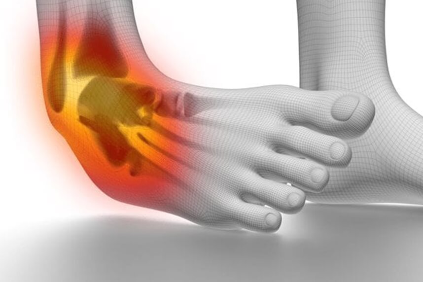
The talocrural joint (ankle) is a joint complex that allows the orientation of the foot in all directions of space, shock absorption, and weight transmission to the support in static and locomotion. To withstand the action of high stresses, the ankle joint must be stable in both joint statics and dynamics. The stability of the ankle is mixed, resulting from the combined action of bone and ligament elements. The geometric conformation of the articular surfaces is mainly responsible for bone stability.
A sprain occurs when the ligaments are partially or completely damaged, following an abnormal movement of this joint. Although ankle sprains are common minor injuries, approximately 25% of those who have an ankle sprain will experience long-term joint pain and instability.
The most common cause of ankle sprain is inversion. If this movement is repeated (tennis, football, basketball) or simply if a patient happens to make several sprains, the ligaments in the lateral area of the ankle (anterior and posterior talofibular and calcaneofibular) can be completely damaged, in which case they must be surgically reconstructed.
.jpg)
Surgical technique
Under spinal anesthesia, an incision of about 2 cm is made in the anteromedial area of the knee and the gracilis muscle is harvested along with its tendon, to be used as a ligament graft.
An incision of about 4 cm in the lateral area of the ankle is made, distal to the peroneal malleolus. The ankle fascia and the articular capsule are highlighted, respectively the bony landmarks: peroneal malleolus, talus neck, and calcaneus.
We dissect the anterior edge of the fibula with the highlight of the abutment remaining from the anterior talofibular ligament and practice 1 channel of 4.5 mm long 25 mm in the talus, and one with the same dimensions in the fibula.
Subsequently, at the lower pole of the fibula, the calcaneal-fibular ligament is highlighted, the abutment remained and the fibula is tunneled with 4.5mm, and the calcaneus with 5.5mm.
.png) | .png) | .png) |
The ligament graft is fixed in the talus and the fibula, with a 5mm bioinert anchor. Subsequently, the ligament graft goes in the heel with a thicker anchor, of 6.5 mm, because the quality of the bone is weaker at this level.
The surgical technique can also be performed arthroscopically, but the patient will have 4 incisions of about 1 cm each at the end. From a surgical point of view, it is a more difficult technique to perform and does not have the same medical accuracy compared to the open technique.
If the lesion is not chronic, the best option is reinserting the anterior talofibular ligament with the help of two 2.8mm titanium rings, and overstabilization by Internal Brace - a thick thread is used, which is fixed and stretched with 2 PEEK bioinert anchors.
Postoperatively
After the intervention, the patient remains hospitalized for 1 day. He will receive pain medication and antibiotics during hospitalization. The operated limb is immobilized in a plaster splint, and the patient is advised not to make ankle movements, 14 days. Free walking is not allowed immediately. You need to use crutches, even if the chosen intervention was minimally invasive.
Patients will wear a compressive bandage on the foot for 5 days. Patients can return to family and professional activities quickly, up to 3-4 weeks, if they have office work, and 10-12 weeks, if they have fieldwork.
At home
Although recovery after this operation is much faster than a classic intervention, it will still take a few weeks for you to fully recover the operated joint. You should expect pain and discomfort for at least a week postoperatively. You can use a special ice pack, which will reduce the pain and inflammation. You must be careful not to lean on the operated area in the first weeks because the pain and discomfort can worsen. You can take a bath, but without wetting the bandage and incisions. The threads are suppressed at 14 days postoperatively.
At 3 months postoperatively, an MRI is necessary to see how the tendon suture heals. Driving is allowed after 6-8 weeks and hard physical work after 10 weeks.
Physical therapy plays a very important role in the rehabilitation program, and the exercises must be followed by a physical therapist until the end of the recovery period.
It is very important to follow the recovery program strictly and seriously for the surgery to be a success. Our medical team works on average with the patient after this intervention, 12-16 weeks until complete recovery of the operated area.
Following any surgery, medical recovery plays an essential role in the social, professional, and family reintegration of the patient. Because we pursue the optimal outcome for each patient entering the clinic, recovery medicine from Centrokinetic is based on a team of experienced physicians and physical therapists and standardized medical protocols.
MAKE AN APPOINTMENT
FOR AN EXAMINATION
See here how you can make an appointment and the location of our clinics.
MAKE AN APPOINTMENT
BUCHAREST TEAM


CLUJ NAPOCA TEAM


BRASOV TEAM


Asiguratori privati

Parteneriate
CENTROKINETIC FLOREASCA
Bucharest, 1st District,
Mircea Eliade Street, no. 18,
Eliade Tower, Entrance A, Floor 3

| Navigation | Explore 3D |
CENTROKINETIC Barbu Vacarescu
Bucharest, 2nd District,
Barbu Vacarescu Street, no. 102,
102 The Address, ground floor.

| Navigation | Explore 3D |
CENTROKINETIC MILITARI
Bucuresti, Sector 6,
Bld. Iuliu Maniu, nr. 15G,
Cladirea PBT, etaj 3.

| Navigation |


CENTROKINETIC CLUJ-NAPOCA
Cluj-Napoca,
Str. Intre Lacuri, nr.1
Intrarea este in spatele cladirii.

| Navigation |


SOCRATES CLINIC TIMISOARA
Timisoara, Calea Martirilor 1989,
nr. 1.
Telefon: 0371 785 374.

| Navigation |
CONTACT
Phone: 0319693
E-mail: programari@centrokinetic.ro
Operating hours:
Monday – Friday: 07:00 – 21:00
Saturday: 09:00 – 14:00
Sunday: closed.
MAKE AN APPOINTMENT

