.jpg)


See details
READ MORE

.jpg)
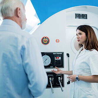
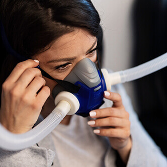
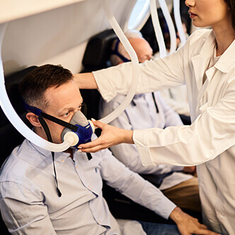
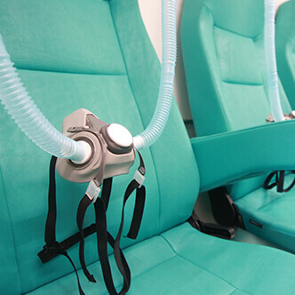
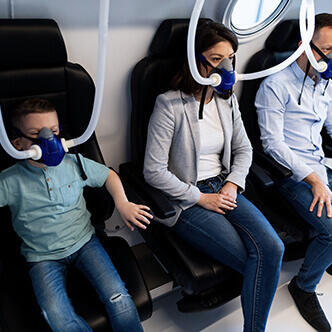
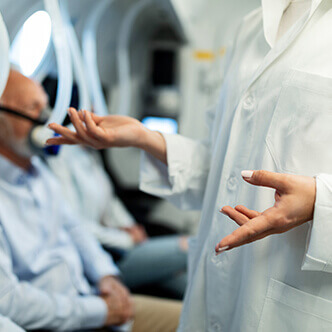
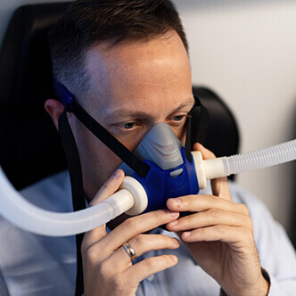
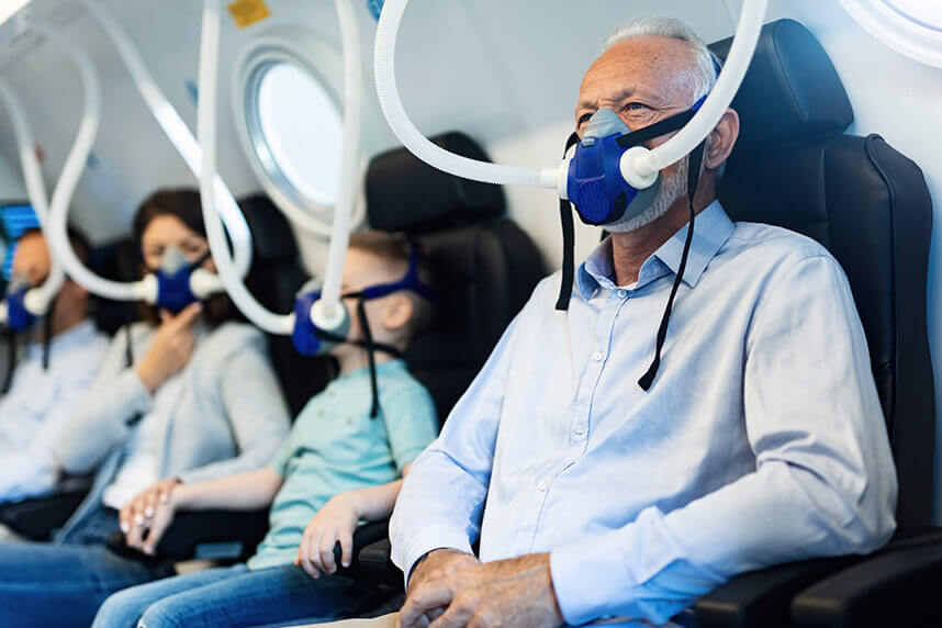
Discover the hyperbaric medicine center opened in our clinic. Centrokinetic has the top-performing hyperbaric chamber in Bucharest, with multiple medical and anti-aging uses. The Baroks chamber has 5 seats, and operates at a constant pressure of 2.5 atmospheres, being fully automated and having protocols for each condition, and can be used individually for each patient.
Patients who use the clinic's hyperbaric therapy services benefit from:
Centrokinetic is keeping contact with prestigious clinics and universities in Belgium, the Netherlands, France, and Greece to constantly update treatments to provide patients with the best medical solutions.
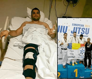
Soft tissue radionecrosis refers to the delayed effects of radiation therapy, i.e long-term damage to the irradiated anatomical area. Radiation can damage capillary beds and arterioles that lead to relative tissue hypoxia, leading to characteristic fibrosis, and these tissue changes can develop over time, away from initial radiation exposure. Soft tissue radionecrosis can develop between 6 months and a few years after exposure. Capillaries can regress through the process of angiogenesis, but in tissue damaged by hypoxic radiation, new capillaries tend to grow disorganized, leading to telangiectasias. This abnormal neovascularization results in improper tissue perfusion that can subsequently cause tissue necrosis and skin ulceration. Only minor trauma or surgical procedures can lead to tissue rupture and ulceration. Radiation can damage superficial and deep tissues. Radionecrosis of the spine and brain can be particularly problematic to treat. The radiation exposure dose is usually over 5000 cGy (Cents-gray) but can be done with up to 3000 cGy.
.jpg) |
|
Soft tissue radionecrosis can occur in any organ or tissue that has received substantial radiation exposure, a medical conduit commonly used to treat cancer. The initial radiation damage (acute phase) in the tissue is in the DNA, and if the damage is severe enough, the cells will not be able to recover. This will lead to cell death. If these are cancer cells, this is the desired effect of the radiation, but when normal tissue that is in the field of ionizing radiation is damaged, then unwanted changes can occur in the tissue.
Initially, there is edema and inflammation in the tissue due to radiation, which leads to clinical erythema. After the radiation-induced erythema, the tissue may develop hypoxia, and fibrous tissue appears.
These changes in fibrotic tissue are a precursor to the development of delayed effects of radiation therapy. Damaged tissue has a lack of normal capillaries and arterioles, growing stroma, which gives the tissue the fibrotic appearance and texture. Such tissue is hypoxic due to the more distant distance of oxygen diffusion from red blood cells that pass through fewer capillaries. Not only the normal capillary beds are missing, but the capillaries present are often disorganized and do not provide adequate tissue perfusion for normal oxygenation. Proliferative endarteritis and fibrosis result in ruptured tissue, either spontaneously or often in combination with trauma and/or infection.
In the United States, there are more than 1.2 million cases of invasive cancer diagnosed annually and about half of these patients will receive radiation therapy to relieve symptoms or treat the disease. About 3% to 5% of patients who have had radiation therapy for cancer will develop delayed effects of radiation therapy, such as soft tissue radionecrosis or problems with wound healing. The risk of developing this problem depends on the amount of radiation delivered, the type of ionizing radiation, and the tissue that has been radiated.
The dose of radiation exposure is usually over 5000 cGy, sometimes at least 3000 cGy. The biological effect of radiation is DNA damage, lipid peroxidation, and protein denaturation. Cells exposed to radiation will either die or malfunction and then undergo cell repair.
The cells that are most affected by radiation are the cells that divide faster, which are found in the dermis, mucosa, and vascular tissue. Endothelial cells are especially prone to damage, which leads to fibrotic changes and the lack of cells that function normally in radiation-damaged tissue. Cellular sensitivity to radiation starts from the most sensitive tumor cells, reaching endothelial cells, fibroblasts, muscles, and then nerves.
The acute clinical phase occurs in the first 6 months after radiation therapy, which is often fractionally dosed. This phase may not involve gross tissue changes but may cause dose-dependent changes. These changes can be manifested clinically by hair loss, erythema, skin diseases, and skin ulcers. Lung tissue can develop radiation-induced pneumonitis, which usually occurs 2 to 3 months after radiation therapy and is considered a subacute phenomenon. Clinically pneumonitis behaves like chronic bronchitis. It can resolve gradually or progress to a chronic problem such as pulmonary fibrosis. If radiation therapy is applied to the spinal cord, then demyelination may occur and, clinically, this condition is known as Lhermitte. Most of the time this symptom will resolve without treatment, but sometimes the symptoms can persist for more than 6 months and will become a chronic condition. These delayed effects of radiation therapy are usually seen more than 6 months after radiation therapy but may occur 5 or more years later. The delayed effects of radiation therapy are dependent on the tissue that has been irradiated.
The pathology behind soft tissue damage follows the principle that cells that have a faster rate of rotation or divide at a higher rate are more sensitive to radiation and are more damaged than cells that don’t undergo division.
Arterioles may try to grow again, but this proliferative endarteritis results in disorganized growth of vascular tissue and often does not provide adequate oxygenation of the tissue to maintain normal function. The relatively hypoxic and fibrotic tissue is now susceptible to rupture resulting in trauma or infection. The resulting chronic wounds are very difficult to treat, and further surgery and grafting can be problematic due to a lack of tissue vascularity.
It is important to have a history about when radiotherapy was given, what type of ionizing radiation was used, and what was the dose of radiation that the tissue received. Also, when the patient has a problematic wound, it is important to consider other factors that can cause poor wound healing, for example, malnutrition, macro-vascular disease, tobacco use, diabetes, age, and repeated tissue trauma. For example, a lesion in the pelvic area may not heal simply by treating the tissue, but also by previous radiation therapy. It is also important what surgical procedures have been performed on the problematic wound.
The acute effects of radiation on soft tissues in the development of erythema, tissue edema, skin pigmentation changes, hair loss, and mucosal ulceration should be studied. The acute effects of radiation therapy are usually self-limiting and are treated with supportive care and the use of antibiotics if the infection develops due to infected skin ulcers.
Usually, the damaged radiated tissue will recover in about a month with reduced inflammation, edema, and repair of damaged endothelial cells. If the arterioles do not recover, then this precipitates the formation of a hypoxic environment with fibrotic changes that can become permanent and can lead to delayed effects of radiation therapy. Delayed effects can cause wounds that are difficult to heal, and the tissue develops progressive endarteritis and an obliterative process of the arterioles that decreases blood supply to the tissue, and the hypoxic environment causes the typical fibrotic change. Clinically, the damaged skin tissue has a contracted appearance, a woody appearance, and a waxy appearance. Telangiectasias are commonly observed on the surface and it is common for this skin to ulcerate with minor or spontaneous trauma.
When evaluating a problematic wound in previously radiated tissue, it is important to determine if the problem is due to soft tissue radionecrosis and not some other cause. Usually, the accumulated radiation dose was over 3000 cGy and more frequently 5000 cGy. For example, a squamous cell carcinoma of the skin previously treated with 1000 cGy would be very unlikely to develop a future skin ulcer from the delayed effects of radiation therapy. More likely, in this scenario, the skin ulcer may be a recurrence of skin cancer and a biopsy should be done.
Wound cultures or bacterial cultures on tissue biopsy may help target antimicrobial therapy and treatments adapted to secondary infections, which are common in soft tissue radionecrosis. It is often difficult to determine whether tissue necrosis results from delayed effects of radiation or side infections. Methicillin-resistant Staphylococcus aureus (MRSA) is particularly problematic due to its pathogenicity and antimicrobial resistance. Therapy should be adapted based on antimicrobial sensitivity.
Not all soft tissue radionecrosis is easily identified by clinical examination. For example, cystitis or hemorrhagic proctitis may be a delayed effect of radiation therapy for pelvic cancers. Evaluation of cystitis and hemorrhagic proctitis will often require cystoscopy or colonoscopy for further diagnosis and treatment. A biopsy may be done during these procedures to confirm the diagnosis and to help look for recurrent cancer or secondary cancer. Often, the diagnosis can be confirmed by CT examination or MRI scan. Spinal cord demyelination in determining the delayed effects of radiation therapy and electrical pain in the lower extremity with the extension of the spine may be characteristic of radionecrosis of the soft tissues of the spine.
Conventional therapies for incurable wounds and bleeding problems, as seen in soft tissue radionecrosis, are unsatisfactory and often unsuccessful in controlling symptoms. Because the tissue lacks adequate vascularity to provide oxygen and nutrients for the tissue to heal, surgery has a higher failure rate and can contribute to further tissue damage and decomposition. Tissue damaged by radiation is also more prone to complications such as infection, especially after surgery.
Hyperbaric oxygen therapy generally has a response rate of about 80%, with an improvement in the patient's symptoms of soft tissue radionecrosis, but the tissue never recovers to what is considered normal tissue. Clinically, what is seen is an improvement in fibro-atrophic tissue changes. For example, improvement in granulation of the injured tissue, reduction or resolution of bleeding in the affected tissue, improvement in osteocyte function, and improvement in neurological symptoms.
Hyperbaric oxygen therapy is useful in treating soft tissue radionecrosis through improved oxygenation of the tissue during treatment, and after 20-30 sessions, angiogenesis is stimulated with the formation of new capillary beds and granulation tissue, which bring a higher blood supply for healing. wounds.
Not only the clinical parameters can be monitored in response to therapy, but improvement in transcutaneous oxygen measurement and fluent angiography may be sought.
Cancer patients who have had a history of bleomycin chemotherapy have an increased risk of pulmonary fibrosis, even if the chemotherapy was given remotely. This has been a relative contraindication, but some doctors will treat the patient if the benefits outweigh the risks. A patient who is actively receiving cis-platinum has a high risk of bladder toxicity if he also receives hyperbaric oxygen therapy and this is a contraindication. Also, a relative contraindication is if the patient had a history of spontaneous pneumothorax with underlying lung conditions with air cushions.
Theoretically, the use of hyperbaric oxygen therapy should be beneficial for angiogenesis and repair of brain radionecrotic tissue.
The differential diagnosis of soft tissue radionecrosis should take into account other causes of tissue necrosis. Necrotizing infections, especially Staphylococcus and Streptococcus infections, can cause tissue necrosis and should be excluded from the assessment of wound infectious disease and deep tissue culture.
Hemorrhagic cystitis is a complication of pelvic radiation, and the delayed effects can be very difficult to treat and often lead to further bladder damage that occurs after cauterization treatments. Proctitis is also a delayed soft tissue radionecrosis that can lead to significant rectal bleeding. Such bleeding problems can be severe enough to require repeated blood transfusion and have significant morbidity.
It is always important when the patient encounters such a situation, the specialist to ensure that there is no recurrence of cancer or a secondary carcinoma or a precancerous adenomatous polyp that causes bleeding, to make all the necessary tests.
Common forms of soft tissue radionecrosis are central nervous system (CNS) radionecrosis (cerebral and spinal cord), radiation-induced cystitis where hemorrhage results, radiation-induced proctitis, vaginal radionecrosis, and laryngeal radionecrosis. The morbidity for these patients is substantial, with 50% of patients having complications if future surgery is required on the radiation field. Annually, there are between 6,000 and 30,000 patients who develop this condition in the United States. Radiation-induced cystitis with bleeding can be ameliorated in approximately 80% of patients, a significant reduction in hematuria being observed.
The beneficial effects of hyperbaric oxygen therapy are sustained effects due to the major angiogenesis that takes place in the tissue, along with the supply of oxygenated blood and improved granulation of the injured tissue. Hyperbaric oxygen therapy is most beneficial when used in combination with good surgical technique, as demonstrated by Dr. Marx's extensive studies on osteoradionecrosis of the mandible and the development of the Marx protocol.
.jpg)
The risk of hyperbaric oxygen therapy techniques includes hypoglycemia in diabetic patients, especially if taking insulin and / or hypoglycemic agents, ear barotrauma, pneumothorax and seizures of toxicity and rarely pulmonary toxicity with oxygen. Cancer patients who have had a history of bleomycin chemotherapy have an increased risk of pulmonary fibrosis, even if the chemotherapy was given remotely. This was a relative contraindication, but some doctors will treat the patient if the benefit outweighs the risk. Due to the hyperbaric environment of the oxygen chamber, there is always the risk of fire, which is why a series of strict protocols are followed to prevent the devices from causing accidents.
The synthesis of collagen that is essential for tissue regeneration is dependent on oxygen and the energy produced by mitochondria through oxidative phosphorylation that forms ATP also comes from the presence of oxygen. Hyperbaric oxygen also has effects on the improved function of neutrophil's ability to kill bacteria and makes neutrophils less adherent to endothelial cells, helping to reduce tissue edema.
The patient with soft tissue radionecrosis must be treated with a multidisciplinary approach with hyperbaric oxygen therapy, and not as an isolated treatment. It is recommended to apply hyperbaric oxygen therapy in combination with appropriate surgical debridement, promptly, with antimicrobial therapy for infection, adequate nutrition, oncological considerations and consideration of plastic surgery options for treating the wound.
The affected patient must receive good wound care with the proper use of dressings to control wound moisture and bacterial growth. Cases can become very complex and not all therapy options are reasonable.
Hyperbaric oxygen therapy is often added as an adjunct especially if the tissue is fibrotic or has continuous progressive necrosis. At this point in therapy, a necrotizing infection present in soft tissue radionecrosis can be treated. It is often beneficial to perform repeated selective debridement of the wound while using hyperbaric oxygen therapy. It is especially useful to have a selective debridement about once a week after the hyperbaric oxygen treatment because the viable tissue still has a healthy color, and the necrotic tissue will stand out well, making selective debridement easier. Negative pressure therapy can be started once the tissue no longer has active tissue necrosis.
Once a healthy bed of granulation tissue has formed, treatment options are either to allow the wound to heal or to consider grafting the skin or closing the flap obtained after surgery. At this stage of the process, a plastic surgeon may help in the treatment and may even use extra treatment with hyperbaric oxygen to efficiently weld the tissues.
Centrokinetic is the place where you will find clear answers and solutions for your motricity problems. The clinic is dedicated to osteoarticular diseases and is divided into the following specialized departments:
Find the latest news by following the Facebook and YouTube accounts of the Centrokinetic clinic.
Diving accidents are accidents that can occur during diving, being specific to this activity, and are always an emergency. Learn the benefits of hyperbaric therapy and why it is useful in diving accidents.
READ MOREHyperbaric oxygen therapy has multiple medical and anti-aging uses. Like any medical treatment, hyperbaric oxygen therapy has certain contraindications. Find out here what are the contraindications for hyperbaric oxygen therapy.
READ MOREScientists in Israel say they can reverse the aging process. The key to this success seems to be hyperbaric oxygen therapy – breathing pure oxygen while sitting in a pressurized chamber for a certain amount of time. In this case, it's 90 minutes/session, with a frequency of 5 sessions per week, for 3 months.
READ MORECystoid or cystic intestinal pneumatosis (intestinal emphysema) is a symptom that can occur in many gastrointestinal diseases. Hyperbaric oxygen therapy can be a successful treatment of cystoid intestinal pneumatosis and granulomatosis with polyangiitis.
READ MOREGynecological cancers treated with a combination of external beam radiation and brachytherapy, especially cervical and vaginal cancers, can result in the apex of the vagina receiving a high dose of radiation. Hyperbaric oxygen therapy has positive effects on the radiated tissues, especially the head, neck, anus, and rectum.
READ MOREHyperbaric oxygen therapy involves the use of so-called levels of oxygen under pressure to increase the level of oxygen in the blood. The use of hyperbaric oxygen therapy involves oxygen treatment for soft tissue radionecrosis. Read this article and find out more.
READ MOREProlonged CO exposure is responsible for more than half of fatal poisonings and is also one of the leading causes of poisoning in Western countries. We aimed to compare the effectiveness of therapy with hyperbaric oxygen (HBO) versus normobaric oxygen (NBO) in the setting of carbon monoxide poisoning (COP).
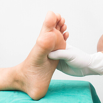
READ MOREHyperbaric oxygen therapy may be effective for Wagner's grade 3 and 4 diabetic foot ulcers and need to study the real problems with patients seeking treatment and demonstrates the need to study the real problems with patients seeking treatment. The results show that it is important to follow the treatment in order for the HBOT to be efficient.
READ MOREHyperbaric oxygen is used in sports medicine to reduce hypoxia and edema and is also effective in treating stroke injuries and acute traumatic peripheral ischemia. When used clinically, hyperbaric oxygen should be considered as an adjuvant therapy used as early as possible after the diagnosis of the lesions.
READ MOREMuscle stretches are the most common muscle injuries suffered during performance sports. Rapid recovery from muscle injury is crucial for elite athletes who regularly are exposed to training and increased competition. Hyperbaric oxygen therapy is a safe and effective method, being a non-invasive treatment
READ MOREHyperbaric therapies are methods used to treat disease or injury using pressures higher than the local atmospheric pressure inside a hyperbaric chamber. The long-term effects are neovascularization (angiogenesis in hypoxic soft tissues), osteoneogenesis, and stimulation of collagen production by fibroblasts. This is beneficial for wound healing and recovery after irradiation.
READ MOREProctitis is the inflammation of the rectal mucosa causing pain, discharge, and other unusual symptoms. Pain can occur during bowel movements, it can be acute or chronic. Symptoms may vary, but the most common is tenesmus (the feeling of needing to go to the toilet), a sensation that persists even after using the toilet. This treatment should be offered to patients who fail to recover with conventional treatments for radiation-induced proctitis.
READ MOREOsteomyelitis is an infection of the bone or marrow caused by bacteria or mycobacteria. Hyperbaric oxygen treatments can be considered an American Heart Association (AHA) Class II recommendation for the treatment of chronic, refractory osteomyelitis
READ MOREHyperbaric oxygenation allows a controlled increase in oxygen pressure in the blood. This technique can be used in cases of tinnitus and sudden deafness, when certain changes in the inner ear and brain generate a lack of oxygen and, therefore, a limited intake of energy.
READ MOREHyperbaric oxygen therapy has been found to ameliorate the damaging effects of reperfusion by early modulation of inflammation, maintenance of metabolic function in downstream tissues, and reintroduction of oxidation scavengers.
READ MOREHBOT has a beneficial effect on burn wound healing by reducing edema and ensuring there is adequate oxygen in microcirculation. It may speed up epithelialization and suppress unnecessary inflammation that could negatively affect normal wound healing. With further research, HBOT may become an adjuvant therapy to surgery.
READ MOREHyperbaric therapy is a form of medical treatment that involves exposing the body to pure oxygen at a higher pressure than normal. There are about 45 diseases approved worldwide to be treated with hyperbaric oxygen.
READ MOREMalignant otitis externa is a rapidly spreading bacterial infection that is aggressive and may be fatal if left untreated. Hyperbaric oxygen therapy (HBOT) is a medical treatment in which the entire body is placed in an airtight chamber at increased atmospheric pressure and has been proven to be effective for several different medical conditions.
READ MOREIf left untreated, MI will lead to the progressive loss of viable cardiomyocytes, impaired heart function, and congestive heart failure. Oxygen cycling therapy serves as a very attractive option for the treatment of myocardial infarction, because it offers some of the greatest benefits while reducing treatment time and inconvenience to the subject.
READ MOREThe present study has demonstrated that adjunct HBOT enhances the reduction of ulcer area and depth at 4 weeks in T2DM patients with ischaemic DFUs. HBOT is known to ensure hyperoxygenation of ischaemic tissue and restoration from hypoxia. Discover the hyperbaric medicine center open in our clinic. Centrokinetic has the top-performing hyperbaric chamber in Bucharest.
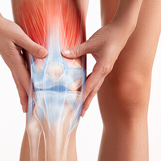
READ MOREOsteonecrosis of the knee (ONK) is a form of aseptic necrosis resulting from ischemia to subchondral bone tissue. Typically, treatment is invasive. Hyperbaric oxygen therapy (HBOT) may provide a noninvasive alternative by improving oxygenation and reperfusion of ischemic areas. This study evaluates the efficacy of HBOT in a series of ONK patients.
READ MOREFemoral head necrosis (FHN), also called avascular necrosis, or femoral head osteonecrosis is a common multifactorial condition that affects patients of any age and can lead to substantial clinical morbidity. Hyperbaric oxygen therapy (HBO) is one of the proposed treatments. Indeed, tissue oxygen promotes angiogenesis that reduces edema. Read about the effectiveness of this treatment.
READ MORECentral retinal artery occlusion (CRAO) is a devastating and common eye condition. It presents a sudden, unilateral, and painless loss of vision. Even when treated promptly, an acute obstruction of the central retinal artery usually leads to severe and permanent loss of vision.
READ MOREThere are numerous studies reported for the effectiveness of HBO in the treatment of osteoradionecrosis of various bone tissues. In addition to its usefulness in treating osteoradionecrosis, this therapy can prevent it. It also combats the negative effect of irradiation, stimulates osseointegration, and improves the survival rate of the implant.
READ MOREThe auditory function in the inner ear is maintained by the cochlea, which is known to have a high oxygen demand. Hyperbaric oxygen can increase the tension of oxygen in the perilymph and restore hearing in a significant number of patients with sudden hearing loss. Patients can be treated in a single-seater hyperbaric chamber or in a multiplace chamber.
READ MORELimb trauma, which leads to direct tissue damage, plus local hypoxic disorders caused by the resulting edema, causes acute peripheral ischemia. Surgical treatment and hyperbaric oxygen are not concurrent treatment modalities but are best used to complement each other in order to provide the best outcome for the patient.
READ MORESmall gas embolisms, as in this case, present serious risks, especially the complication of cerebral air embolism. To prevent neurological complications, it is necessary to urgently remove the air bubble. HBOT reduces the volume of the bubble, helps eliminate nitrogen, and improves the oxygenation of potentially hypoxic tissue. See the results of hyperbaric therapy in venous embolism.
READ MOREOsteoradionecrosis (ORN) is a common consequence of radiation provided to cancer patients. Currently, hyperbaric oxygen therapy (HBOT) has a major role in improving wound healing in patients with ORN.
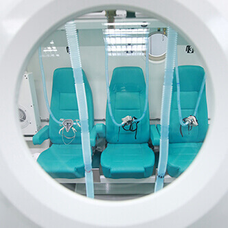
READ MOREDiscover the hyperbaric medicine center open in our clinic. Centrokinetic has the top-performing hyperbaric chamber in Bucharest, with multiple medical and anti-aging uses. The Baroks chamber has 5 seats, and operates at a constant pressure of 2.5 atmospheres, being fully automated and having protocols for each condition, and can be used individually for each patient.
READ MORESee details
READ MORESee details
READ MORESee details
READ MORESee here how you can make an appointment and the location of our clinics.

CENTROKINETIC FLOREASCA
Bucharest, 1st District,
Mircea Eliade Street, no. 18,
Eliade Tower, Entrance A, Floor 3
| Navigation | Explore 3D |
CENTROKINETIC Barbu Vacarescu
Bucharest, 2nd District,
Barbu Vacarescu Street, no. 102,
102 The Address, ground floor.
| Navigation | Explore 3D |
CENTROKINETIC MILITARI
Bucuresti, Sector 6,
Bld. Iuliu Maniu, nr. 15G,
Cladirea PBT, etaj 3.
| Navigation |
CENTROKINETIC CLUJ-NAPOCA
Cluj-Napoca,
Str. Intre Lacuri, nr.1
Intrarea este in spatele cladirii.
| Navigation |
SOCRATES CLINIC TIMISOARA
Timisoara, Calea Martirilor 1989,
nr. 1.
Telefon: 0371 785 374.
| Navigation |
Phone: 0319693
E-mail: programari@centrokinetic.ro
Operating hours:
Monday – Friday: 07:00 – 21:00
Saturday: 09:00 – 14:00
Sunday: closed.
MAKE AN APPOINTMENT