
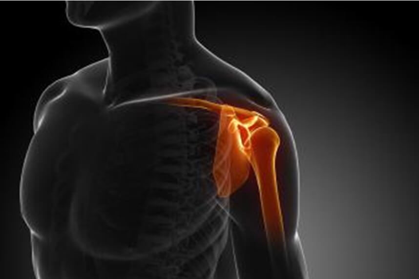
The shoulder complex is made up of 3 bones: the humerus (arm bone), the clavicle, and the scapula (shoulder blade), which are interconnected by ligaments, tendons, and muscles, and 4 joints: gleno-humeral, acromio-clavicular, scapulo-thoracic, and sterno-clavicular. It is a very complex joint and, at the same time, the most mobile joint in the body, allowing a wide range of movements.
The humeral head is ball-shaped and partially fits into a cavity called the glenoid. The glenoid cavity is much smaller in relation to the humeral head, allowing the remarkable mobility found at this level. Stability is achieved through the connection of ligaments and the glenoid labrum, a special type of cartilage that increases the articular surface area and creates better stability in the joint.
The humerus remains close to the glenoid cavity thanks to the joint capsule and the stabilizing action of the strong rotator cuff muscles that surround it. Above the humeral head is a bony prominence that completes the scapular spine, called the acromion. Under the acromion, muscles forming the rotator cuff are inserted. In this space, the shoulder tendons pass over each other during shoulder movement.
The efficiency of these movements is achieved due to the presence of bursae (fluid-filled sacs located at the joints). The subacromial bursa is the largest in this region. Besides providing stability, the bursae facilitate the lifting and rotation of the arm. The two biceps tendons and the pectoralis major muscle insert near the humeral head. This entire complex is covered by the deltoid muscle.
.jpg)
The shoulder is the most mobile joint in the body. The price paid for this extraordinary mobility is stability. Thus, the shoulder is the most mobile and at the same time the most unstable joint in the human body, which often makes it subject to dislocations. When one of the structures that stabilize the shoulder is damaged after trauma or weakened due to a personal configuration associated with genetics, some movements of the upper limb cause abnormal and inconsistent sliding of the humeral head, which causes pain and a feeling of instability that reaches its peak in cases of shoulder dislocation. In such cases, there is immediate functional impairment accompanied by intense pain. Reducing the dislocation (repositioning the humeral head in the glenoid cavity) is possible through specific maneuvers that are not easy to perform. To confirm the diagnosis, the doctor will suggest a CT or MRI to determine the appropriate therapeutic approach for your case. The joint will be immobilized with a bandage/brace for about 3 weeks, followed by a rehabilitation period. Rehabilitation itself plays an important role because the use of the upper limb requires a free and painless joint, and the most common problem that occurs after a dislocation episode is permanent instability that sooner or later recurs. Initially, the shoulder will be stiff and painful with hypertrophic musculature, which usually causes discomfort or worry/fear in the patient. Our duty is to personalize the physiotherapy program to achieve the delicate balance that allows a wide range of normal amplitude movements with the greatest possible stability.
.jpg)
This injury consists of a complete or incomplete tear of the acromio-clavicular ligament that holds the acromion and clavicle together. The consequence is an upward dislocation, associated with pain and inability to move the shoulder. These joint injuries are direct (a direct fall, cycling, football, a traumatic contact) with the face down. Symptoms are characterized by local pain and deformation of the acromio-clavicular profile. Clinical conditions are variable, depending on the characteristics of the injury. Radiological examination provides specific information for the acromio-clavicular joint. Ultrasound examination can also reveal ligament injuries and provide information about the level of dislocation. MRI is suggested in cases of other suspected injuries, such as those of the rotator cuff. Treatment is usually conservative and involves immobilizing the joint for at least 20 days. This solution allows the injured structures to heal in the best position, although a period of rehabilitation is necessary after removing the bandage to restore movement and regain strength, which are inevitably compromised by the trauma itself and also by prolonged immobilization. If the injury is complex, surgery is mandatory to keep the two joint ends together.
.jpg)
Clavicle fractures are among the most common causes of bone injuries. The fracture is most often located at the junction between the medial and lateral thirds (80%), this being the most vulnerable point. When the fracture is complete, the medial fragment moves upward due to the action of the sternocleidomastoid (SCM) muscle, while the lateral fragment moves downward due to the weight of the arm and the action of the deltoid. The traumatic mechanism occurs especially due to falls on an outstretched arm (fall from a horse, motorcycle, or bicycle). The pain at the fractured site can be intense enough to make arm movement impossible. The area is swollen and deformed at the edges of the fractures, altering the normal shoulder profile. The injury is diagnosed with the help of X-rays, but sometimes muscle tissue ultrasound is sufficient. However, it is important to perform a CT scan for fractures that are difficult to diagnose, while an MRI is suggested when there is suspicion of rotator cuff injury. Fracture healing depends on the alignment conditions of the bone fragments. This requires the shoulder to be held back by a functional bandage in an "8" shape, which is necessary, even if uncomfortable. The clavicle usually heals in 3 weeks, but if the fracture is more complex, immobilization will be prolonged. When the bandage is removed, a bone callus can be seen under the skin, as if it were a step. Physiotherapy is extremely important: when the bandage is removed, hydrotherapy sessions and physiotherapy sessions should begin to restore the range of shoulder motion as quickly as possible. Other important objectives in recovery are restoring muscle strength and neuromotor control of the shoulder.
.jpg)
These fractures usually occur at the surgical neck of the humerus. In people over 50, a simple fall can cause this type of fracture. In younger individuals, humerus fractures occur following more violent trauma: falling from a height, car accidents, or sports injuries. Symptoms are characterized by intense pain that extends beyond the injury site and immediate functional impairment. The pain worsens even after minor shoulder movements. X-rays are usually sufficient for diagnosis. When the fracture is small, treatment is conservative associated with limb immobilization with a compressive bandage for 20-25 days and a cycle of physiotherapy: mobilization and controlled pendulum exercises are performed to increase the range of motion, and proprioception exercises are added for muscle strength.
.jpg)
The term SLAP lesion most often represents an anterosuperior lesion of the glenoid labrum associated with the detachment of the long head of the biceps tendon. This injury occurs after a violent movement in throwing sports or a fall on the shoulder. The onset of symptoms is very rarely due to a defined trauma. Usually, the medical history refers to various traumatic episodes during overhead activities. Very often you will be able to reproduce the voluntary dislocation movement, usually occurring in a throwing position. You will likely feel pain when resting, which tends to worsen when performing overhead movements. The pain is accompanied by a sensation of ligamentous laxity and joint noises. In mild cases, surgery can be avoided by completely avoiding sports or overhead movements that tend to worsen the injury. Physiotherapy aims to strengthen the internal rotator muscles, restoring the full range of motion as quickly as possible without forcing external rotation. Stretching the capsule and posterior cuff can alleviate symptoms.
.jpg)
Rotator cuff tears can be partial or complete and are very common. In 90% of cases, these injuries are degenerative, with only 10% resulting from trauma. Degenerative tears are the natural progression of chronic rotator cuff tendinopathy and external subacromial impingement syndrome. Traumatic tears are associated with younger patients, sports enthusiasts who get injured while performing athletic activities, or are the consequences of a shoulder fall in case of injury, usually associated with anterior dislocations of the scapulo-humeral joint or humerus fractures. Treatment is usually conservative when dealing with partial tears, with continuous check-ups that allow the patient’s autonomy to be preserved for independence in daily life. Complete tears, however, have a somewhat more difficult prognosis, and surgery in most cases is the only solution, which also applies to partial tears that do not respond to conventional treatment after a few months.
.jpg)
A tear of the long head of the biceps often involves the proximal portion (the insertion area on the scapula). It is more common in people over 40 who suffer from chronic pain caused by impingement syndrome. This leads to chronic tendon suffering in cases of overload and degeneration. The tear usually occurs after intense effort and is associated with older individuals with rotator cuff suffering. The pain is sudden, localized, and violent, generally accompanied by a sound similar to an elastic break, and the appearance of an anterior bulge. The diagnosis is clinically established. X-rays can be helpful for patients suffering from chronic shoulder pain due to rotator cuff injury. The treatment usually used for such injuries is conservative and goes through all 5 phases of rehabilitation. An aesthetic deformity tends to remain after the tear, but full elbow bending and strength recovery are nonetheless achievable.
.jpg)
Adhesive capsulitis or frozen shoulder is a condition caused by specific inflammation of the shoulder joint capsule. It can be idiopathic (without a known cause) or due to joint injuries or surgical interventions performed at this level, especially if treated with extended immobilization. It is characterized by the progressive loss of range of motion due to the loss of flexibility of the joint capsule.
This injury is characteristic of women aged between 40-60 years and patients with clinical signs of depression. Motor difficulties are especially encountered at the level of both internal and external rotation. A radiological examination by a specialist is recommended to identify the presence of calcifications or other pathologies. The recovery process is usually quite slow and in some cases can last more than 2 years. We can help you regain your range of motion as quickly as possible through specific therapeutic exercises, hydrotherapy, and active physiotherapy using wireless electrotherapy or TECAR WINBACK. The doctor may recommend managing the issue even with medication.
If after 12 months of rehabilitation there are no successes, mobilization under anesthesia or arthrolysis (surgical intervention aimed at restoring joint mobility by cutting the ligaments and the capsule surrounding the joint) may be suggested.
.jpg)
When we talk about shoulder instability, all traumatic pathologies that can occur at this level should be considered, such as dislocations, luxations, subluxations, and pathological joint laxity. Various classifications have been proposed, but we will refer to instability involving patients with signs of congenital pathological joint laxity, associated with instability at the shoulder level.
Instability can also affect athletes who use the maximum range of motion at this joint, such as gymnasts, volleyball players, weightlifters, or swimmers.
The traumatic effect is produced by repetitive overhead movements which, due to joint weakening, simultaneously cause mechanical anomalies to the nervous structure and periarticular soft tissues (repeated microtrauma) which cause pain.
If you recognize yourself in the above description, it means that you most likely started to feel shoulder pain or suffer from sensory disturbances (paresthesias) like "dead arm" or numbness of the upper arm when performing daily or sports activities. You may have encountered various occasions of dislocation or subluxation without significant trauma.
Conservative treatment consists of trying to manage this complex medical situation. This work is largely completed by improving the biomechanics of the joint by toning the muscles that stabilize the joint. In particular, in sports that have high arm movements, it is necessary to strengthen all the rotator cuff muscles, as they are involved in controlling fine movements and stabilizing the humeral head. Recovery of neuromuscular control is essential, as coordination deficit is typical. Coordination exercises can be an alternative in field rehabilitation, where patients will be subjected to dynamic and more specific activities.
If after at least 6 months of conservative therapy there are no qualitative results, a surgical intervention and continuation of postoperative physical therapy may be necessary.
.jpg)
Every time the upper limb is raised above the head, there is a narrowing of the space between the head of the humerus and the acromion. In this subacromial space, the tendons of the rotator cuff are located, which are protected by the subacromial bursa. High-performance sports, or activities that require repetitive overhead movements, rotator cuff muscle imbalance, or the irregular character of the acromion can cause increased interaction within this space. These movements create a conflict between the soft structures (subacromial bursa) and the acromion and can lead to the formation of calcium deposits on the lower part of the subacromial space. Pain usually occurs at night. Due to the pain, we begin to avoid using the arm, and this causes the appearance of intra-articular adhesions and worsens the medical condition.
A correct and rapid diagnosis followed by an early start of physical therapy is the key in this type of condition to stop the vicious cycle mentioned above. An early approach to the condition makes the prolonged need for anti-inflammatory drugs decrease and also prevents the occurrence of side effects caused by adopting analgesic postures. If investigations demonstrate the presence of relevant anatomical changes, such as near-complete tearing of one or more rotator cuff tendons or the appearance of significant calcifications, surgical intervention followed by physical therapy may be necessary.
.jpg)
A proximal humerus fracture occurs mostly (80% of cases) in women suffering from osteoporosis.
The type of treatment suggested in most cases is surgical and consists of stabilizing the fragments with Kirschner wires or Rush pins. When osteosynthesis is desired, with screws and plates, or in patients over 40 years old or in the case of 4-part fractures, even with prosthetics.
Regardless of the type of surgical intervention suffered, immediate mobilization of the limb is essential to prevent post-traumatic stiffness. Passive mobilization begins as soon as possible, in a pain-free zone. We will always maintain contact with the surgeon to establish together the timing and method of joint recovery.
You will probably frequently undergo check-up x-rays to verify the maintenance and stability of the fracture. An MRI scan may be necessary to verify the integrity of the humeral head which can progress to necrosis in these cases. Starting from the third week post-surgery we begin active exercises with reduced amplitude, we are interested in re-establishing a balance at the scapula level and for this reason, we will perform elevation movements at the scapula level, paying special attention to rotation movements. Pendulum exercises will be prescribed along with exercises for activating scapular stability and manual treatment for the scapulohumeral girdle.
Water recovery (hydrokinetotherapy) is also extremely important since it helps muscle relaxation. Starting from the 6th postoperative week, the physical therapy program will aim to increase mobility even more. The introduction of active exercises into the protocol for the rotator cuff will be with the surgeon's approval to avoid fracture disassembly, which is possible in the first months post-operation.
The treatment period requires approximately 3-4 months of therapy to recover the entire range of motion and for complete functionality.
(2).jpg)
The surgical repair of the injury can be performed either through classical surgery or arthroscopy. In this case, the damage to the periarticular tissue is significant, and recovery is less difficult. The surgeon's goal is to re-stabilize and correct the tendon insertion areas.
When the injury is old, retraction and degeneration of tendon fibers can prevent the surgeon from performing an anatomical reattachment. In this case, specialists will aim to reunite the edges of the injury. The more correct the suture is, the greater the chances of success. For this, a double suture can even be performed for even more significant strengthening of the insertions.
Collaboration with the surgeon during the recovery process is extremely important. They will provide us with information about the tissue characteristics, the degree of tension applied to the sutures, and the specifics of the surgical intervention.
The recovery program must be properly structured and monitored. Throughout physical therapy, we consider two important aspects: respecting the biological recovery time of the suture and ensuring an early stimulus for regaining the full range of motion.
(2).png)
Surgical treatment involves repositioning the glenoid labrum by anchoring the detached part of the glenoid ligaments and, if necessary, suturing the long head of the biceps brachii. This may or may not be associated with an intervention on the rotator cuff.
The surgical operation can be performed through arthroscopy or classical surgery. After the intervention, it is recommended to use a brace that will keep the arm in slight abduction (slightly away from the trunk) and neutral rotation for 3-4 weeks. Between the third and sixth postoperative weeks, a gradual recovery of passive mobility at all levels should be possible. From this point on, physical therapy will aim to increase the range of motion to achieve maximum functionality within 10-12 weeks post-surgery. Starting from week 3, isometric training begins in a comfortable position, with special attention to exercises that stimulate the long head of the biceps.
However, active mobility is maintained below 90 degrees until the sixth postoperative week. For athletes, the recovery program will be more intensive and accelerated, aiming for full recovery of movements within 6-8 weeks post-surgery (if the shoulder is not immediately immobilized, there is a high risk of developing a frozen shoulder with a significant deficit in external rotation).
See here how you can make an appointment and the location of our clinics.

CENTROKINETIC FLOREASCA
Bucharest, 1st District,
Mircea Eliade Street, no. 18,
Eliade Tower, Entrance A, Floor 3
| Navigation | Explore 3D |
CENTROKINETIC Barbu Vacarescu
Bucharest, 2nd District,
Barbu Vacarescu Street, no. 102,
102 The Address, ground floor.
| Navigation | Explore 3D |
CENTROKINETIC MILITARI
Bucuresti, Sector 6,
Bld. Iuliu Maniu, nr. 15G,
Cladirea PBT, etaj 3.
| Navigation |
CENTROKINETIC CLUJ-NAPOCA
Cluj-Napoca,
Str. Intre Lacuri, nr.1
Intrarea este in spatele cladirii.
| Navigation |
SOCRATES CLINIC TIMISOARA
Timisoara, Calea Martirilor 1989,
nr. 1.
Telefon: 0371 785 374.
| Navigation |
Phone: 0319693
E-mail: programari@centrokinetic.ro
Operating hours:
Monday – Friday: 07:00 – 21:00
Saturday: 09:00 – 14:00
Sunday: closed.
MAKE AN APPOINTMENT


