
Soft tissue ultrasound is performed on the recommendation of the attending physician in cases where this non-invasive ultrasound investigation method is considered more efficient or cheaper than a conventional radiography, a CT scan, or an MRI. Below you will find a complete guide, including information about everything this type of ultrasound entails, when it is recommended, how it is performed, how long it lasts, how it is interpreted, and how to prepare for a soft tissue ultrasound.
Soft tissue ultrasound, or soft tissue echography, is a type of non-invasive imaging investigation that provides detailed images of superficial soft tissues such as skin, dermis, and subcutaneous soft tissues, as well as superficial anatomical structures such as ligaments, tendons, salivary glands, thyroid, and lymph nodes.
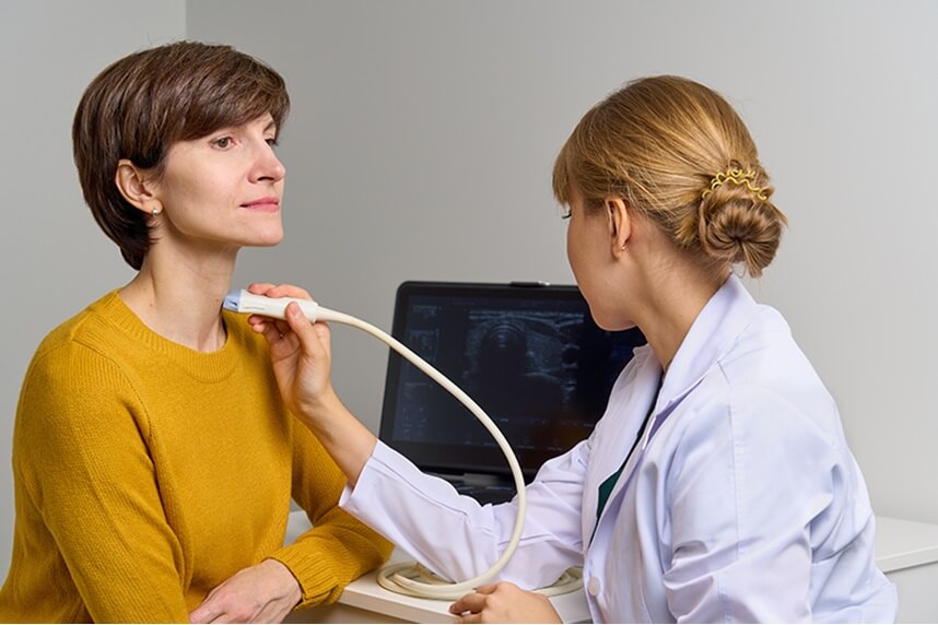
Soft tissue ultrasound is performed with a linear transducer with a high working frequency that is applied to the skin of the area to be examined, displaying images of the examined soft tissues on the ultrasound device's monitor. A transparent gel is applied to the examined skin area to facilitate the sliding and efficiency of the ultrasound. Soft tissue ultrasound is usually performed on the following anatomical areas:
Usually, the attending physician recommends a soft tissue ultrasound to establish a diagnosis or for the evaluation and monitoring of a soft tissue condition for which the patient is already undergoing treatment.
Soft tissue ultrasound is recommended in the following cases:
In most cases, no special preparation is required before a soft tissue ultrasound. However, depending on the area to be examined, the doctor may ask the patient not to eat or drink before the procedure.
It is preferable for the patient to wear loose and comfortable clothing, preferably with a blouse or shirt that can be easily lifted or removed to allow access to the area to be examined.
Also, wearing jewelry or other metal objects, such as piercings, on the area to be examined is not recommended, as they can interfere with the ultrasound waves, resulting in erroneous ultrasound images.
Last but not least, the patient should inform the doctor or medical staff of any details from their medical history that may be relevant and ask the doctor or medical staff about any specific instructions they should follow.
Soft tissue ultrasound is a non-invasive medical procedure that uses ultrasound waves to create images of the body's soft tissues. The exact procedure may vary depending on the area to be examined and the doctor's instructions. Here is how this procedure is usually conducted:
The patient is positioned in a way that allows easy access to the area to be examined. For example, if the abdomen is being examined, the patient may be lying on their back or side. The next step is applying ultrasound gel to the skin in the area to be examined. This gel helps transmit the ultrasound waves and improves the quality of the images. The doctor uses a handheld probe called a transducer, which emits ultrasound waves into the respective area. The transducer is gently moved over the skin, and the ultrasound waves are reflected by the soft tissues and captured by the transducer. The captured ultrasound waves are transformed into real-time images on a monitor. The doctor analyzes these images to evaluate the structures and soft tissues of the examined area. The procedure may be repeated in different positions or angles to obtain more detailed images or to examine different parts of the area. After the procedure is completed, the ultrasound gel is removed from the skin.
The results of the soft tissue ultrasound are given immediately. Their accuracy depends on the quality of the ultrasound equipment used and the experience and skills of the doctor performing the ultrasound, who interprets the images obtained in the context of the patient's medical history. Also, if the patient has had previous ultrasounds, the doctor compares the images. Any change is an indication of the disease's progression.
When analyzing the ultrasound images, the doctor pays particular attention to the following aspects:
How long does a soft tissue ultrasound take?
The duration of a soft tissue ultrasound varies depending on the area being investigated and the complexity of the case. In general, a soft tissue ultrasound can take between 15 and 45 minutes.
Is soft tissue ultrasound painful?
Soft tissue ultrasound is a non-invasive procedure that involves using an ultrasound probe to create images of the body's soft tissues. Therefore, it is painless. The probe is moved over the skin covered with gel, which facilitates sliding and reduces any possible discomfort. Applying light pressure during the procedure is normal. If the pressure causes acute pain, the patient should signal this to the doctor.
When are additional ultrasounds or other tests needed after a soft tissue ultrasound?
The specialist doctor interpreting the images obtained during the ultrasound may request another ultrasound or additional tests if:
Is soft tissue ultrasound suitable for all age groups?
Being a non-invasive imaging technique without ionizing radiation, soft tissue ultrasound is suitable for all age groups, from infants to seniors.
Being a non-invasive procedure, soft tissue ultrasound poses no risks for any age group. On the contrary, it is the preferred imaging procedure because:
Given that the accuracy of the result of a soft tissue ultrasound depends equally on the quality of the ultrasound equipment and the skills of the doctor, it is very important for patients to be directed to imaging centers that can offer both quality equipment and adequately trained doctors who can interpret the images accurately.
See here how you can make an appointment and the location of our clinics.


CENTROKINETIC FLOREASCA
Bucharest, 1st District,
Mircea Eliade Street, no. 18,
Eliade Tower, Entrance A, Floor 3
| Navigation | Explore 3D |
CENTROKINETIC Barbu Vacarescu
Bucharest, 2nd District,
Barbu Vacarescu Street, no. 102,
102 The Address, ground floor.
| Navigation | Explore 3D |
CENTROKINETIC MILITARI
Bucuresti, Sector 6,
Bld. Iuliu Maniu, nr. 15G,
Cladirea PBT, etaj 3.
| Navigation |
CENTROKINETIC CLUJ-NAPOCA
Cluj-Napoca,
Str. Intre Lacuri, nr.1
Intrarea este in spatele cladirii.
| Navigation |
SOCRATES CLINIC TIMISOARA
Timisoara, Calea Martirilor 1989,
nr. 1.
Telefon: 0371 785 374.
| Navigation |
Phone: 0319693
E-mail: programari@centrokinetic.ro
Operating hours:
Monday – Friday: 07:00 – 21:00
Saturday: 09:00 – 14:00
Sunday: closed.
MAKE AN APPOINTMENT


