
VALG FLAT FOOT CORRECTION TECHNIQUE WITH HYPROCURE
Discover the open MRI imaging center in our clinic. Centrokinetic has a state-of-the-art MRI machine, dedicated to musculoskeletal conditions, in the upper and lower limbs. The MRI machine is open so that people suffering from claustrophobia can do this investigation. The examination duration is, on average, 20 minutes.
Centrokinetic attaches great importance to the entire medical act: investigations necessary for correct diagnosis (ultrasound, MRI), surgery, and postoperative recovery.
The talocrural joint (ankle) is a joint complex that allows the foot movement in all directions of space, shock absorption, and weight transmission to the support in static and locomotion. To withstand the action of high stresses, the ankle joint must be stable in both joint statics and dynamics. The stability of the ankle is mixed, resulting from the combined action of bone and ligament elements. The geometric conformation of the articular surfaces is mainly responsible for bone stability.
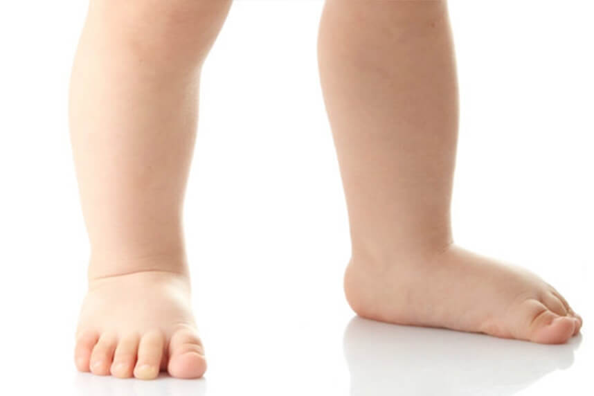
Pes valgus represents a deformation of the ankle and hindfoot, clinically easy to recognize, which consists of decreasing the height of the medial longitudinal arch. However, there is no clinical or radiological definition, unanimously accepted, of the average height, of the values considered normal of the medial longitudinal arch of the foot.
The height of the medial longitudinal arch is determined by the shape of the bones and the strength of the ligament structures. The muscles intervene only for balance because it has been shown that the leg muscles are inactive during orthostatism (standing).
.jpg)
Pes valgus occurs at the time of loading and represents a change in normal bone ratios at the ankle, so the heel is rotated externally and flexed dorsally relative to the talus, so there is a pronation movement, valgus of the foot. These changes in interosseous relationships create a collapse of the medial longitudinal arch.
Surgical technique
The surgical treatment consists in blocking the hyper pronation movement of the foot, valgus, and collapse of the medial longitudinal arch with the help of a special implant, which is inserted at the level of the talocalcaneal joint, in the tarsal sinus.
.jpg)
It is a simple surgery (incision 1 cm, minimal risks) in terms of operative amplitude (minimum incision, short duration, extremely fast recovery), is performed under radiological control, and the results are remarkable.
Under spinal (spinal) anesthesia, an incision of about 1 cm is made in the lateral part of the ankle, at 2 cm anteroinferior to the tip of the peroneal ankle. The subcutaneous tissue is dissected and an anatomical space, called the tarsi sinus, is highlighted. With the help of surgical scissors, the talocalcaneal ligament is sectioned and a guide pin is inserted.
Subsequently, the diameter of the tarsal sinus canal is measured, and the Hyprocure implant is inserted under radiological control.
.jpg)
This implant completely corrects the deformation of the ankle and offers patients a normal life, without pain and without repercussions over time.
The history of this surgery began 15 years ago in the USA, where Dr. Michael E. Graham invented an implant that once inserted between the heel and the talus, blocks the hyper pronation movement of the ankle, and corrects the deformity. The 15-year history has led to the use of this implant in patients aged between 3 and 94 years, placing over 30,000 implants worldwide. The failure rate of the intervention is only 6%, but even in these cases in which the implant was removed after a few years, the ankle remained in an acceptable degree of correction, and the patients were satisfied.
.jpg)
Postoperatively
After the operation, the patient remains hospitalized for 1 day. He will receive pain medication and antibiotics during his hospitalization. The operated limb is not immobilized in a plaster splint, and the patient is advised to make ankle movements starting the day after the intervention. Walking with a load is not allowed immediately, you need to use crutches, even if the chosen intervention was minimally invasive.
Patients will wear a compressive bandage on the foot for 5 days. Patients can return to daily activities quickly, up to 3-4 weeks, if they have office work, and 10-12 weeks, if they have fieldwork.
At home
Although recovery after this operation is much faster than a classic intervention, it will still take a few weeks for you to fully recover the operated joint. You should expect pain and discomfort for at least a week postoperatively. You can use a special ice pack, which will reduce the pain and inflammation. You must be careful not to lean on the operated area in the first weeks because the pain and discomfort can worsen. You can take a bath, but without wetting the bandage and incisions. The threads are suppressed at 14 days postoperatively. At 6 weeks postoperatively, an MRI is necessary to see how the tendon suture heals. Driving is allowed after 8 weeks, and hard physical work after 10-12 weeks.
Physical therapy plays a very important role in the rehabilitation program, and the exercises must be followed by a physical therapist until the end of the recovery period.
It is very important to follow the recovery program strictly and seriously for the surgery to be a success. Our medical team works on average with the patient after this intervention, 12-16 weeks until complete recovery of the operated area.
Following any surgery, medical recovery plays an essential role in the social, professional, and family reintegration of the patient. Because we pursue the optimal outcome for each patient entering the clinic, recovery medicine from Centrokinetic is based on a team of experienced physicians and physical therapists and standardized medical protocols.
MAKE AN APPOINTMENT
FOR AN EXAMINATION
See here how you can make an appointment and the location of our clinics.
MAKE AN APPOINTMENT
BUCHAREST TEAM


CLUJ NAPOCA TEAM


BRASOV TEAM


Asiguratori privati

Parteneriate
CENTROKINETIC FLOREASCA
Bucharest, 1st District,
Mircea Eliade Street, no. 18,
Eliade Tower, Entrance A, Floor 3

| Navigation | Explore 3D |
CENTROKINETIC Barbu Vacarescu
Bucharest, 2nd District,
Barbu Vacarescu Street, no. 102,
102 The Address, ground floor.

| Navigation | Explore 3D |
CENTROKINETIC MILITARI
Bucuresti, Sector 6,
Bld. Iuliu Maniu, nr. 15G,
Cladirea PBT, etaj 3.

| Navigation |


CENTROKINETIC CLUJ-NAPOCA
Cluj-Napoca,
Str. Intre Lacuri, nr.1
Intrarea este in spatele cladirii.

| Navigation |


SOCRATES CLINIC TIMISOARA
Timisoara, Calea Martirilor 1989,
nr. 1.
Telefon: 0371 785 374.

| Navigation |
CONTACT
Phone: 0319693
E-mail: programari@centrokinetic.ro
Operating hours:
Monday – Friday: 07:00 – 21:00
Saturday: 09:00 – 14:00
Sunday: closed.
MAKE AN APPOINTMENT

