Actualizat: 18-03-2025 / Publicat: 11-06-2020
For all traumatic or chronic diseases of the musculoskeletal system, the Centrokinetic private clinic in Bucharest is prepared with an integrated Orthopedic Department, which offers all the necessary services to the patient, from diagnosis to complete recovery.
The Department of Orthopedic Surgery of Centrokinetic is dedicated to providing excellent patient care and exceptional education for young physicians in the fields of orthopedic surgery and musculoskeletal medicine.
Centrokinetic attaches great importance to the entire medical act: investigations necessary for correct diagnosis (ultrasound, MRI), surgery, and postoperative recovery.
Discover the open MRI imaging center in our clinic. Centrokinetic has a state-of-the-art MRI machine, dedicated to musculoskeletal conditions, in the upper and lower limbs. The MRI machine is open so that people suffering from claustrophobia can do this investigation. The examination duration is, on average, 20 minutes.
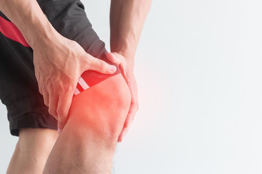
Anterior cruciate ligament rupture (ACL) is the most common ligament injury of the knee, causing over 50,000 ligament reconstructions per year in the United States. The risk of this injury has increased greatly among the average population, due to increased participation in sports activities.
Most of the time, the rupture (ACL is due to a rotational mechanism. The repercussions of the ACL rupture must be very well understood because the rupture of the anterior cruciate ligament has no consequences on the flexion-extension movements of the knee. The patient completely regains this movement after 4-6 weeks of physical therapy and does not feel instability when performing this movement (eg knee bends). Instead, the knee is vulnerable to rotational and torsional movements: the patient feels instability in rotational movements of the body with the foot locked to the ground (pivoting sports), but also in everyday life when performing these rotational movements. Clinical examination of the patient is very important to detect these instabilities: sagittal and rotational. Rotational stability is provided by ACL and ALL (anterolateral ligament of the knee), there are situations in which both ligaments rupture and rotational instability are very high. That is why the clinical examination and the patient's anamnesis (detailed description of the accident) are very important.
The surgical technique is an extremely controversial problem, which has undergone many changes in the last 3 decades, and currently, anatomical reconstruction of the ligament is opted for. This reconstruction aims to position the graft as close as possible to the natural ligament, but this idea is difficult to achieve because the natural ligament has both an origin and an insertion, wider than the graft used; it has a double orientation towards the graft and it has 2 strips with a well-defined position and roles, compared to the graft which is a single strip. Therefore, this surgery fails to fully reach the desired parameters, ideal in terms of graft positioning.
![i.php?p=p7(3).jpg]()
There are many methods of reconstructing the LIA, among which we mention:
- Autograft harvested from the patient is the most commonly used graft, it is cheap, it is very good because it integrates best, but it does not always have the desired dimensions: length and thickness. The autograft is of several types: hamstrings, patellar tendon (BTB), quadriceps tendon, fascia lata. Currently, the most commonly used as the first intention is the hamstrings type, and the BTB type is used in revisions (when the initial graft is also broken). However, a category of athletes in whom the BTB graft is recommended as the first intention is footballers. In the USA, for high-performance athletes (soccer, American football, even basketball, although this category due to repeated jumps, patellar tendinopathy occurs later), the graft used is BTB.
- Allograft (obtained from suppliers specialized in sports medicine): it is an ideal graft in terms of size and structure, being an Achilles tendon. The graft has the desired thickness, the tendon is very resistant, but the chances of integration are lower than in the case of autografts. That is why this graft, in the world, is much less used, being an option only in certain cases.
- Artificial graft (graft made of synthetic material, used in performance athletes, because it ensures a quick return to sport)
.jpg)
Recent studies show that ACL reconstruction restores knee stability. Initially, ACL reconstruction was an open, visual, invasive procedure, and the results, despite restoring joint stability, were poor due to the many complications that occurred: flexion contracture, patellofemoral pain, decreased mobility, quadriceps muscle atrophy. But technology has advanced, with minimally invasive methods appearing, and the results are commensurate.
A very important role in the process of obtaining a good surgical and functional result is the choice of the operative moment. Initially, it was established that ligamentoplasty should be done as soon as possible after injury, so as not to cause intraarticular fibrosis, which would lead to decreased mobility. Subsequent studies have shown a higher rate of arthrofibrosis in patients operated on in the first week after injury, compared to patients operated on after 4-6 weeks. Then, it was shown that a correct, firm, and complete recovery program equals the results of the two groups. However, the success of LIA reconstruction requires preoperatively: complete hyperextension, the disappearance of inflammatory phenomena, good muscle tone, maintaining knee mobility.
Surgical technique (with hamstrings graft)
Make a vertical incision of 2-3 cm in the inferomedial area of the knee, 2 cm medial to the tibial tubercle. The fascia of the sartorius muscle is carefully dissected, the two muscles are identified by fascia: semitendinosus and gracilis. At this level, the fascia is incised, just above them, with great attention to the medial collateral ligament, and the 2 muscles are identified. They are carefully dissected, the fibrosis around the two tendons is removed. Then, the tendons are disinserted and the semitendinosus muscle tendon is initially harvested. Depending on its size, the tendon of the gracilis is also harvested.
Subsequently, the knee joint is arthroscopically inspected and the integrity of all intra-articular tissues (menisci, cruciate ligaments, articular cartilage) is monitored.
Preparation of the femoral canal: it is done with the knee in hyperflexion, with the help of a special guide, which is applied on the posterior cortex of the external femoral code. Insert a guide brooch that is removed through the skin, then the drill with a diameter of 4.5 mm until it pierces the outer cortex and the length of the canal is measured with a dipstick. Depending on this length, the following landmarks can be established: the length of the endobutton necessary to fix the femoral part of the graft, the length on which the 4.5mm canal widens. The final width of the canal depends on the thickness of the graft (number by number). Currently, there are also adjustable endobuttons that simplify the surgical technique.
Currently, the femoral canal can be made retrograde, less invasive, with the help of a device called FlipCutter, which is a brooch that bends at the tip once it has been inserted into the joint with a special guide.
The preparation of the tibial canal depends on the modulus that fixes the graft:
- If the fixation involves the use of an interference screw, then the preparation is done with a special guide through which a brooch is inserted into the joint cavity. By knee extension, it is checked if there is an impingement with the femoral condyle or the posterior cruciate ligament. Depending on the distal thickness of the graft, a drill (number by number) is used to create the tibial tunnel to the level of the joint surface.
- If the fixation involves the use of an adjustable endobutton (all inside technique), then we use a retrograde brooch, and the tunnel will have a diameter equal to that of the graft. Our medical team has been performing the all-inside technique for about 4 years, with excellent results. The advantages are the increased contact of the graft with the bone, by the lack of the screw, which gives much greater chances of graft integration.
After fixing the graft, the anteroposterior and latero-lateral stability of the knee must be checked, as well as the rotational instability. Depending on the result obtained, one can also opt for the restoration of the anterolateral ligament of the knee (ALL). If we need to reconstruct the anterior cruciate ligament after a failure of the initial intervention, we opt for the patellar or quadriceps tendon. Reconstruction with the patellar tendon is a technique used extremely frequently in the past, but a method left in the background today, being used more in revisions or in performance athletes. The operative technique involves harvesting the patellar tendon, 1/3 average together with 2 bone pellets from the patella and the tuberosity of the tibia. This tendon will represent the new graft, currently fixed with end buttons or screws. This intervention has excellent results on instability, but has the disadvantages of slower recovery due to postoperative pain, but also a higher rate of intra- or postoperative complications. The advantage is the faster integration of the graft, the bone-tendon-bone interface being the most favorable.
Post surgery
After the intervention, the patient remains hospitalized for 1-2 days. He will receive pain medication and antibiotics during his hospitalization. The operated limb is partially immobilized in a mobile knee splint.
At home
Although recovery from an osteotomy is much faster than a classic intervention, it will still take a few months for you to fully recover your knee joint. You should expect pain and discomfort for at least a week postoperatively. Ice will reduce pain and inflammation.
You must be careful not to sleep on the operated knee in the first weeks because the pain and discomfort can worsen. You can take a bath, but without wetting the bandage and incisions. The threads are suppressed at 14 days postoperatively.
Physical therapy plays a very important role in the rehabilitation program, and the exercises must be followed by a physical therapist until the end of the recovery period. It is very important to follow the recovery program strictly and seriously for the surgery to be a success. Our medical team works on average with the patient after this intervention, 18-24 weeks until complete recovery of the knee.
Following any surgery, medical recovery plays an essential role in the social, professional, and family reintegration of the patient. Because we pursue the optimal outcome for each patient entering the clinic, recovery medicine from Centrokinetic is based on a team of experienced physicians and physical therapists and standardized medical protocols.

.jpg)
.jpg)
.jpg)

.jpg)
.jpg)


