
Discover the open MRI imaging center in our clinic. Centrokinetic has a state-of-the-art MRI machine, dedicated to musculoskeletal conditions, in the upper and lower limbs. The MRI machine is open so that people suffering from claustrophobia can do this investigation. The examination duration is, on average, 20 minutes.
Centrokinetic attaches great importance to the entire medical act: investigations necessary for correct diagnosis (ultrasound, MRI), surgery, and postoperative recovery.
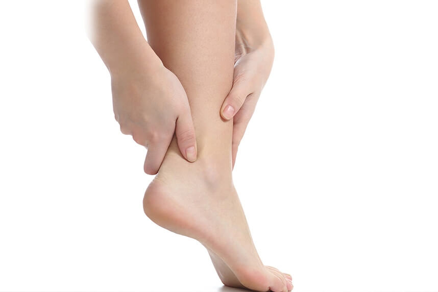
The talocrural joint (ankle) is a joint complex that allows the foot movement in all directions of space, shock absorption, and weight transmission to the support in static and locomotion. To withstand the action of high stresses, the ankle joint must be stable in both joint statics and dynamics. The stability of the ankle is mixed, resulting from the combined action of bone and ligament elements. The geometric conformation of the articular surfaces is mainly responsible for bone stability.
The tendons of the long and short peroneal muscles have their origin near the knee joint and are inserted in the midfoot area, having a role in the stability of the ankle and the internal and external rotational movements.
The two muscles become tendons above the ankle and cross this anatomical area, through the posterior area of the fibula, through a bony groove, being stabilized in this position by a fascia, called the peroneal sheath. The short peroneal tendon is located directly in contact with the fibula and is generally more prone to injury. The long fibula tendon is located behind the short tendon and helps to perform the eversion movement and also to flex the lower leg.
.jpg)
Subluxation or dislocation of the peroneal tendons usually occurs as a result of an associated ligament injury. The peroneal retina, which holds the ligaments in place in the posterior area of the fibula, ruptures, this injury subsequently allowing these structures to dislocate from their normal position. Untreated, this condition will cause inflammation of the tendons and their rupture over time.
.jpg)
The lateral ligaments of the ankle (anterior talofibular ligament (ATFL) and calcaneofibular ligament (CFL)) are located just below and in front of the fibula ankle. Their damage in case of a sprain can cause dislocation of the tendons of the peroneal muscles which can occur immediately or in time.
.jpg)
Surgical technique
This operation can be performed under spinal anesthesia (spinal anesthesia). The patient is placed in lateral decubitus (on one side), with support placed under the ipsilateral leg to allow free movement of the ankle during surgery and easy access to the peroneal tendons and posterior fibula. An incision of 4-6 cm is made along the lateral edge of the fibula and curved distally near the tip of the fibula, according to the anatomical trajectory of the tendons of the peroneal muscles.
Careful dissection highlights the sural nerve posteriorly. The upper retina of the extensor muscles is then incised 1-2 mm posterior to its fibular insertion, to allow easy repair after the procedure. It can also be removed directly from the bone if it is chosen to use a bone reinsertion technique. Often, a marginal "Bankart" lesion is found on the lateral edge of the distal fibula, being created or perpetuated by dislocated peroneal tendons. The rest of the upper retina is carefully preserved.
The peroneal tendons are dislocated from the retromalleolar groove, to be inspected and repaired in case of concomitant injuries and to evaluate the anatomy and fibular cartilage along the posterior aspect of the fibula. The tip of the fibula is exposed, later, and a brooch is inserted retrogradely through the peroneal malleolus, its position can be verified by performing an intraoperative radiograph.
Sequentially, the fibula is tunneled with drills of 4mm, up to 6mm. Repeating intraoperative radiographs may also be useful in determining whether a properly sized drill was used. Subsequently, two corticotomies (bone incisions) are performed on either side of the fibula joint. Next, the articular surface is slightly pushed into the previously formed canal, thus forming a new bony sans. After repositioning the peroneal tendons, their stability is checked and the superior retinaculum of the extensors is repaired.
Postoperatively
After the intervention, the patient remains hospitalized for 1 day. He will receive pain medication and antibiotics during his hospitalization. The operated limb is immobilized in a plaster splint, and the patient is advised not to make ankle movements for 14 days. Charging is not allowed immediately, you need to use crutches, even if the chosen intervention was minimally invasive. Patients will wear a compressive bandage on the leg for 5 days. Patients can return to daily activities quickly, up to 3-4 weeks, if they have office work, or 10-12 weeks if they have fieldwork.
At home
Although recovery after this operation is much faster than a classic intervention, it will still take a few weeks for you to fully recover the operated joint. You should expect pain and discomfort for at least a week postoperatively. You can use a special ice pack, which will reduce the pain and inflammation. You must be careful not to lean on the operated area in the first weeks because the pain and discomfort can worsen. You can take a bath, but without wetting the bandage and incisions. The threads are suppressed at 14 days postoperatively. At 3 months postoperatively, an MRI is necessary to see how the tendon suture heals. Driving is allowed after 8 weeks, and hard physical work after 10-12 weeks.
Physical therapy plays a very important role in the rehabilitation program, and the exercises must be followed by a physical therapist until the end of the recovery period.
It is very important to follow the recovery program strictly and seriously for the surgery to be a success. Our medical team works on average with the patient after this intervention, 12-16 weeks until complete recovery of the knee.
Following any surgery, medical recovery plays an essential role in the social, professional, and family reintegration of the patient. Because we pursue the optimal outcome for each patient entering the clinic, recovery medicine from Centrokinetic is based on a team of experienced physicians and physical therapists and standardized medical protocols.
See here how you can make an appointment and the location of our clinics.

CENTROKINETIC FLOREASCA
Bucharest, 1st District,
Mircea Eliade Street, no. 18,
Eliade Tower, Entrance A, Floor 3
| Navigation | Explore 3D |
CENTROKINETIC Barbu Vacarescu
Bucharest, 2nd District,
Barbu Vacarescu Street, no. 102,
102 The Address, ground floor.
| Navigation | Explore 3D |
CENTROKINETIC MILITARI
Bucuresti, Sector 6,
Bld. Iuliu Maniu, nr. 15G,
Cladirea PBT, etaj 3.
| Navigation |
CENTROKINETIC CLUJ-NAPOCA
Cluj-Napoca,
Str. Intre Lacuri, nr.1
Intrarea este in spatele cladirii.
| Navigation |
SOCRATES CLINIC TIMISOARA
Timisoara, Calea Martirilor 1989,
nr. 1.
Telefon: 0371 785 374.
| Navigation |
Phone: 0319693
E-mail: programari@centrokinetic.ro
Operating hours:
Monday – Friday: 07:00 – 21:00
Saturday: 09:00 – 14:00
Sunday: closed.
MAKE AN APPOINTMENT


