Knee
The knee is composed of the femur at its upper part, which articulates with the tibial plateau at its lower part. Between these two large bones, we have the menisci (two for each knee) which serve to cushion the forces that reach this level. In addition to these large bones, in front of the knee, we have the patella (kneecap) which prevents the knee from extending too much, and laterally we have the fibula.
The knee joint is supported by four strong ligaments: the anterior cruciate ligament (ACL), the posterior cruciate ligament (PCL), the medial collateral ligament (MCL), and the lateral collateral ligament (LCL), which stabilize all movements of the knee. They are assisted in stabilizing the joint by strong muscles (quadriceps, hamstrings, gastrocnemius), but due to the complexity of the joint, knee injuries are among the most common sports injuries.
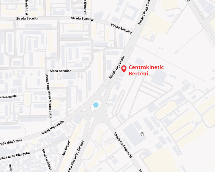
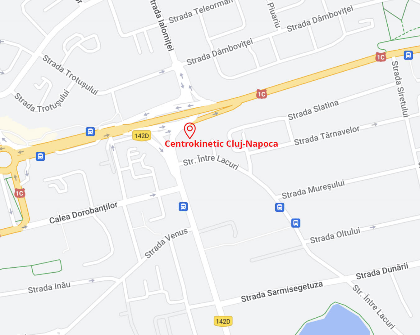
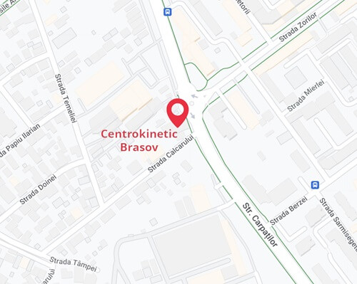
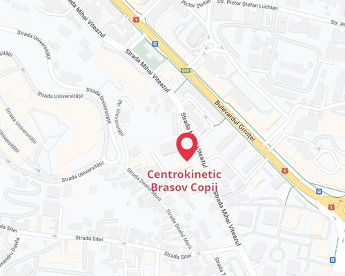
If you suffer from a knee injury or a sports trauma, it can be challenging to find the specialized treatment you need. Our clinic is easy to find and has experts in knee treatment.
We have physiotherapists with extensive experience in treating common and chronic knee injuries, who can help with pain relief, improving the range of motion, and resuming usual or sports activities.
Contents:
- Common Conditions
- patellar dislocation
- knee sprain and dislocation
- tibial plateau fracture
- patella fracture
- anterior cruciate ligament injury
- posterior cruciate ligament injury
- cartilage injuries
- medial/lateral collateral ligament injuries
- meniscus injuries
- patellar and quadriceps tendon rupture
- Chronic Conditions
- osteoarthritis
- iliotibial band syndrome
- patellofemoral syndrome
- patellar tendinopathy or “jumper’s knee”
- Surgical Interventions
- cartilage surgery
- meniscus surgery
- patella surgery
- osteotomy
- knee replacement
- patellar realignment
- posterior cruciate ligament reconstruction (PCL)
- anterior cruciate ligament reconstruction (ACL)
- quadriceps reattachment
- collateral ligament repair
Common Conditions
- Patellar Dislocation
.jpg)
This is a common knee injury either due to a sports trauma or a congenital predisposition.
After excessive stretching of the joint, instability occurs, with a feeling that “something is out of place” as well as pain, inflammation, and difficulty in movement.
If it can be treated conservatively, we use muscle strengthening to increase strength and reduce pain. In cases where surgery is necessary (usually arthroscopy), we have a postoperative rehabilitation program designed to get you back in shape and prevent a new injury.
- Knee Sprain and Dislocation
.jpg)
Knee sprains make up a large percentage of knee injuries, particularly among soccer players, basketball players, volleyball players, skiers, but can also occur in traffic accidents.
Most people who suffer such an injury present with knee inflammation and/or joint pain, with our sports doctors being experts in treating them. Knee dislocations additionally involve ligament or capsular tears that create major instability in the knee. Depending on the extent of the damage, they can be categorized into three different grades: mild, moderate, or severe.
If you have suffered a sprain or suspect a dislocation of the knee, try to apply ice packs as soon as possible and schedule a consultation with a specialist to receive an accurate diagnosis.
- Tibial Plateau Fracture
.jpg)
These injuries are frequently observed in sports, especially skiing.
Patients usually report pain, swelling, and limited knee movement, similar to LCL injuries, though without the associated instability.
Surgery is very rarely recommended in this situation, with a period of immobilization followed by a sufficiently long sports rehabilitation program to treat knee pain, allowing the patient to lead a normal life.
- Patella Fracture
.jpg)
With this type of trauma, pain, inflammation, and limitation of knee movement appear immediately.
Conservative treatment usually starts with immobilization, followed by hydrotherapy and rehabilitation exercises aimed at reducing pain through muscle strengthening.
Surgical intervention may be necessary if the patella is fragmented or dislocated. In this case, our postoperative program helps you recover completely within approximately 3 months.
- Anterior Cruciate Ligament Injury
.jpg)
We frequently encounter anterior cruciate ligament injuries, and for this reason, we can offer significant support in stabilizing the knee without surgery.
Usually, these injuries manifest as intense pain, inflammation, knee instability, and limited joint movement, symptoms for which our center specializes in treatment.
In cases where medical recovery is not sufficient, surgical intervention is necessary to stabilize the injury and restore knee function.
- Posterior Cruciate Ligament Injury
.jpg)
Posterior cruciate ligament injuries are rarer than anterior cruciate ligament injuries and occur mostly due to traffic accidents. Symptoms are also less severe, with pain and knee inflammation possibly appearing later after the injury, making diagnosis more difficult.
- Cartilage Injuries
.jpg)
Joint cartilage problems are common due to repeated incorrect movements or repeated trauma. Severe erosion of the cartilage, known as chondropathy, causes poor gliding of the bone ends. This results in pain, inflammation, and difficulty in movement. If you have been diagnosed with serious cartilage degeneration in a joint such as the knee, you should be aware that recovery can be a very long process.
After a clinical examination, the doctor will prescribe an MRI that can identify the location and extent of the injury.
Four stages of chondropathy have been identified, with increasing severity, which influences the therapeutic approach: conservative treatment may be used for mild injuries, while surgery is usually suitable for more severe stages.
The goal is always to stabilize the joint to stop the vicious cycle leading to joint degeneration. The rehabilitation program will be determined based on the location and degree of the injury. The intention is to alleviate pain, restore tone, and endurance of specific muscle groups that play a crucial protective role.
.jpg)
These types of injuries are usually the result of excessive leg extension following a strong impact.
The severity of the symptoms experienced is directly related to the intensity of the trauma, the degree of ligament rupture, determining the intensity of pain, inflammation, and knee instability.
Treatment also depends on the severity of the trauma, and there are situations where surgical intervention cannot be avoided. However, we can help you recover through hydrotherapy, physiotherapy, and kinesitherapy in a dynamic manner aimed at regaining mobility and stability and returning to a normal lifestyle.
.jpg)
Meniscus injuries cover a wide range of knee traumas and can cause different types of pain depending on the cause. Sports traumas usually cause sharp pain in the knee, while injuries due to everyday movements or cartilage degeneration are generally associated with dull pain triggered by certain movements.
Typically, the specialist doctor will recommend an MRI or CT scan to observe what is happening at the musculoskeletal level before starting a rehabilitation program based on kinesitherapy, physiotherapy, and hydrotherapy for treating the knee and its causes.
This is an extremely severe condition, particularly affecting athletes and those with degenerative tendinitis. Ruptures can be partial or complete, though all are accompanied by severe knee pain, swelling, and difficulties in extending the joint.
At Kinetic Plus, we treat partial ruptures conservatively, primarily by reducing pain and swelling, then increasing muscle strength, before ultimately restoring the patient to full range of motion.
Again, in surgical cases, in collaboration with an orthopedic specialist surgeon, we plan an effective postoperative rehabilitation program to get you back on your feet.
Chronic Conditions
- Osteoarthritis
Osteoarthritis is a very common issue affecting the knee, especially in overweight and female patients. Symptoms are very consistent - knee pain, swelling, a feeling of stiffness, and noises in the joint. The clinical diagnosis is made by the specialist doctor following physical and imaging examinations.
Our kinesitherapy programs focus on pain reduction and increasing the range of motion to improve the overall quality of life for the patient. Weight loss, avoiding excessive strain, and maintaining correct posture are very important.
- Iliotibial Band Syndrome
Iliotibial band syndrome refers to chronic inflammation affecting the iliotibial band of the fascia lata and is caused by persistent mechanical friction.
This condition is particularly common among soccer players, runners, and cyclists and often results from overuse, and training on hard surfaces or uneven terrain. Anatomical peculiarities can play an important role in the development of such pathology.
Knee in varus combined with knee hyperextension can cause excessive tension on the posterior muscle chain, thereby worsening symptoms.
Soft tissue ultrasound is useful in diagnosing this injury, and an MRI can differentiate between this pathology and an external meniscus issue.
For this condition, surgical intervention is recommended to be avoided except in very severe cases. To aid rehabilitation, we will employ a combined approach of manual therapies and kinesitherapy.
- Patellofemoral Syndrome
This condition causes pain felt in the back of the knee and, in severe cases, can lead to patellar dislocation.
The knee is an extremely complex joint, and a slight alteration in the form or function of a muscle group – in this case, the quadriceps – can create increased pressure on the patella, leading to pain, sprain, or even dislocation if the patella moves out of place.
This condition can generally be treated through kinesitherapy in most cases. When the patient has experienced repeated patellar dislocations, surgical intervention is required for fixation. Our proposed kinesitherapy program aims to rebalance the musculo-ligamentous structure of the knee and increase its resistance, so it can handle daily demands.
If surgical intervention is necessary, the specialist will recommend a postoperative medical recovery program during which we will help you increase muscle strength and joint mobility.
- Patellar Tendinopathy or “Jumper's Knee”
This is a very common injury among those who practice sports with frequent, explosive movements, such as basketball and athletics, and is usually caused by overuse or repeated microtraumas.
Swelling (edema) and pain are typically observed around the knee. Knee pain progresses through four clinical stages, starting with mild pain and increasing to severe pain before the tendon eventually ruptures.
Ultrasound is commonly used to observe any tendinopathies.
In most cases, this can be treated without surgery, though the recovery period depends on the severity of the injury. Our therapeutic approach has a very high success rate in recovering from this type of injury.
It most frequently occurs in adolescents who have grown rapidly; this condition causes abnormal stress on the cartilage, leading to repeated microtraumas.
Pain is usually localized around the knee and is exacerbated during physical activity until rest. Knee swelling is also frequently observed.
This condition can resolve on its own once growth is complete, but in the meantime, we can manage symptoms and reduce pain through kinesitherapy treatment.
Surgical Interventions
- Cartilage Surgery
.png)
The surgical techniques used in this case are numerous and varied: some aim to stimulate the remaining cartilage tissue's ability to self-repair through the production of fibrocartilage, while others focus on regenerating new cartilage from scratch and replacing it with new hyaline cartilage. Clearly, the chosen option depends on the severity of the injury, with more severe cases requiring more radical interventions.
Cartilage Erosion, or “cartilage shaving”: this is a procedure that smooths the surface of damaged cartilage. In first-degree lesions, cartilage begins to crumble and forms fibrils, which are removed using a specific tool in an attempt to eliminate the free edges that mechanically conflict with the joint. Long-term results of this strategy are poor. This technique itself is not a final solution as it has no reparative or regenerative capacity and is used only to alleviate symptoms.
Microfractures: this technique uses small needles to create numerous perforations at the subchondral level, spaced 3-4 mm apart. This leads to drawing blood from the bone layer under the cartilage, forming a new layer of inferior quality cartilage (fibrous cartilage) compared to the original (hyaline cartilage), though the new layer is acceptable from a biomechanical standpoint. This intervention is reparative. Loading is generally acceptable one month after surgery, but sports with high impact potential should be avoided for up to 6 months afterward.
Autologous Osteochondral Grafting (Mosaicplasty): a core of cartilage tissue is extracted along with a portion of subchondral bone from non-weight-bearing joints and then inserted over the damaged cartilage, which is properly prepared. This way, the defective cartilage is filled with hyaline cartilage, giving good results even in the long term. This intervention includes a non-weight-bearing period of 30-45 days post-surgery and allows a return to high-impact sports after approximately 8 months.
Autologous Chondrocyte Transplantation: this method involves two distinct operations. First, chondrocytes are harvested from the joint and “cultivated” for a month; after 30 days, the chondrocytes are grafted onto a three-dimensional matrix (hyaluronic acid, collagen, and alginate) and reinserted into the joint to fill the cartilage defect. Long-term results are excellent, but the postoperative recovery time is very long. This intervention includes a non-weight-bearing period of 30-45 days post-surgery and allows a return to high-impact sports after approximately 10 months.
Biomimetic Reconstruction (MaioRegen): one of the most recent advancements in surgery, this technique involves the implantation of synthetic support structures made of collagen fibers and hydroxyapatite. The technique involves a single operation during which the structure is shaped over the defective cartilage. This part is then inserted after allowing the affected surface to bleed so that the totipotent cells contained in the blood can colonize the structure and produce chondrocytes. This intervention includes a non-weight-bearing period of 45-60 days post-surgery and allows a return to high-impact sports after at least 10 months.
Autologous Mesenchymal Cell Transplant: stem cells are harvested from the patient's bone marrow, extracted from the iliac crest. These cells are inserted into a supporting structure loaded with additional growth enzymes extracted from the patient's blood. Finally, this compound is implanted at the site of the injury, filling the cartilage defect. Results from these operations are comparable to those obtained from biomimetic reconstructions, though the initial non-weight-bearing period is shorter, at 30-45 days, while returning to high-impact sports is permitted after at least 12 months.
- Meniscus Surgery
.jpg)
These arthroscopic interventions are performed only after extremely severe injuries or when conservative treatments have failed. There are four major categories of procedures: meniscus suturing, selective meniscectomy, artificial meniscus implantation, and meniscus implantation from a donor (allograft).
Meniscus Suturing: if the size and location of the injury permit, the surgeon will repair the meniscus using a suture. Recovery in this case takes much longer than with simpler medial meniscectomy.
Selective Meniscectomy: when meniscus suturing is not possible, the surgeon will need to remove the torn meniscus fragments to restore the meniscus profile to normal. This type of intervention usually involves a very short hospital stay.
Artificial Meniscus Implantation: this surgical procedure was preceded by a long evaluation period. It is now an extremely popular technique because it yields very good results. A synthetic meniscus prosthesis is introduced arthroscopically, encouraging the development of meniscal tissue and thus delaying the onset of osteoarthritis.
Donor Meniscus Implantation, or Allograft: this surgical technique involves implanting meniscal tissue from a donor, which is then sutured into the recipient's knee. Similar to the artificial meniscus implant, the biological integration period of the new tissue must be closely monitored, and the recovery period will be much longer than for simple meniscectomy.
Regardless of the type of surgical intervention used, postoperative recovery plans include intensive physiotherapy, aquatherapy to allow patients to exercise with reduced weight, and field recovery to regain specific skills.
- Patellar Surgery
.jpg)
This is reserved for severe fractures or cases where fragment dislocation has occurred. This procedure uses osteosynthesis materials mounted on metal frames, although in some cases, total patellectomy (removal of the patella) may be necessary.
In any case, postoperative recovery plans start with continuous passive motion (CPM), followed by physiotherapy starting in the second week after surgery, and then introducing loading two weeks later. Complete recovery from this type of surgical intervention usually takes about three months.
- Osteotomy
.jpg)
Osteotomy for correcting limb deformities is one of the oldest orthopedic procedures in medicine, with the first recorded operation taking place in 1875 on the tibia. It was previously performed to correct knee deformities at all levels, including both hallux varus, but today its use is more restricted. It is now more commonly performed as external tibial osteotomy to correct varus deformity or medial femoral osteotomy to correct valgus deformity.
Various fixation methods can be used, including staples or a plate with screws; in these cases, recovery time can be accelerated due to greater stability of the wound.
Once the stitches are removed, rehabilitation in the pool (hydrokinesiotherapy) can begin. After a visit from an orthopedic specialist and an X-ray, work in our kinesitherapy room can commence.
Post-surgical rehabilitation here aims to increase the patient's independence in daily activities and enable them to run again and resume sports training.
- Knee Replacement
.jpg)
This surgery is recommended in cases of severe knee osteoarthritis where pain is persistent and movement is severely affected, and where diagnostic X-rays have confirmed the severity.
Generally, this type of intervention is recommended for individuals over 60 years old due to the prosthesis's lifespan and the fact that as age increases, the demand for physical performance decreases. Knee replacement surgery should be postponed as long as possible for patients who continue to maintain sufficient functionality and have tolerable pain. In cases where osteoarthritis affects a young person's joint, procedures such as osteotomy are preferred as they correct load axes, reducing pressure on load-bearing joints.
When helping patients with their postoperative recovery, our primary goal is to restore the full range of motion, then muscle strength, and finally coordination through a personalized program of hydrokinesiotherapy and kinesitherapy. With this approach, we typically manage to get patients on their feet about 10-15 days after surgery and on the path to complete recovery.
- Patellar Realignment
.jpg)
There are three surgeries usually considered here:
Lateral Release: A small section of the lateral collateral ligament is removed and reinserted arthroscopically into the patella to restore the normal profile of the kneecap. The effectiveness of this treatment has been debated; however, the intervention is relatively aggressive and allows for a very rapid postoperative recovery.
Distal Realignment: This option is typically chosen in cases of evident misalignment that leads to recurrent patellar dislocations. After surgery, a period of partial immobilization for four weeks is required.
Proximal Realignment: This treatment is often chosen for adolescents, where the medial vastus is harvested to avoid damaging immature bones. The immobilization period required after this surgery ranges from 4-6 weeks. We are proficient in providing post-surgical rehabilitation techniques, regardless of the knee surgeries performed.
Most patients will start in our kinesitherapy room for rehabilitation shortly after surgery, with or without a prosthesis. Proper kinesitherapy is actually more important than the surgery itself in these cases. Most patients should expect to be back to normal after about three months.
- Posterior Cruciate Ligament (PCL) Reconstruction
(2).jpg)
Although the posterior cruciate ligament is rarely operated on in isolation, interventions for it are more common when there are injuries to other ligaments as well. Grafts taken from the gracilis tissue are common in PCL cases, similar to ACL. Bandages are used to immobilize the knee after surgery to maximize natural recovery and healing.
Postoperative recovery will begin with careful recovery of flexion movements for about 3 months before allowing free joint movement. Strengthening the quadriceps muscle through gym training, using electrical stimulation, followed by active exercise, constitutes the initial phase of the recovery process. Recovery after this type of knee intervention generally takes about five months, with hydrokinetotherapy, physiotherapy, and field recovery all playing an essential role.
- Anterior Cruciate Ligament (ACL) Reconstruction
(2).jpg)
Three surgical techniques are used to treat these knee injuries:
- Reconstruction using the semitendinosus (gracilis) tendon
- Reconstruction using the patellar tendon
- Reconstruction using an allograft (donor tendon)
Reconstruction with the Semitendinosus Tendon
Reconstruction with the semitendinosus or gracilis tendon is one of the most commonly used options. This surgical procedure involves using pieces from the tendons of these medial flexor muscles, which are harvested and passed through a bone tunnel into the joint, usually arthroscopically.
In the kinesitherapy program, it is important to consider the time needed for the healing of the muscles used in the surgery.
Reconstruction using the Patellar Tendon
Reconstruction using the patellar tendon involves removing the central third of the patellar tendon through an incision approximately 5 cm in length. This tendon is then introduced into the joint through a bone tunnel under arthroscopic guidance. This type of intervention tends to weaken the knee extensor mechanism, which can lead to painful tendinitis of the quadriceps muscle and the patellar tendon if excessive weights are used during recovery—thus increasing the recovery time and making this option less popular.
Reconstruction with Allograft
Reconstruction with an allograft uses a graft obtained from a donor, such as the Achilles tendon or patellar tendon. This intervention has the advantage of not using the patient's tendons, thus avoiding weakening of the thigh or quadriceps flexor muscles, as seen in the other two options.
Using a bandage for immobilizing the knee after surgery is at the discretion of the orthopedic team. In most cases, crutches are recommended for approximately 3 weeks.
Recovery should start two days after surgery, either in the hospital or at home, before beginning at the recovery center, about ten days later. Recovery from these types of interventions can take up to five months, with recovery activities alternating between hydrokinetotherapy, physiotherapy, and field recovery.
Post-surgical rehabilitation will follow the path dictated by the surgical technique used. It may vary significantly, but in all cases, we will go through the five phases of rehabilitation: starting with reducing postoperative pain and swelling, then recovering normal movement, followed by regaining muscle strength and endurance, then restoring neuromuscular coordination, and finally returning to sports. The first phase of rehabilitation will be conducted by alternating time between the pool and the kinesitherapy room. This moment is particularly delicate as the replaced tissue is vulnerable to mechanical stimuli. If weight is appropriately dosed, it can stimulate the integration and maturation of the new tissue; otherwise, excessive loading can cause catastrophic damage to the implant.
Manual and physical therapies will be alternated in the kinesitherapy room, and we will use proprioception and strengthening exercises according to the specific program established by the kinesitherapist. Hydrokinetotherapy aims to help recover walking and correct movements in the operated joint. If the patient is an athlete, specific exercises in deep water may also be introduced to start reintroducing correct movements before loading the joint.
In the water, as in the kinesitherapy room, resistance and coordination exercises are performed using progressive resistance. In the following months, the patient will start running and performing preparatory exercises for reintroduction into daily activities. During rehabilitation, eccentric training sessions are included, and for performance athletes, when the operated limb is not significantly weaker than the non-operated one, field work begins, provided that the appropriate recovery time for the biological recovery of the operated ligament has passed. In this phase, a threshold test is used to evaluate the condition of the limb and provide the rehabilitation coach with accurate information about the patient's progress so they can develop the most personalized and effective work program. This step-by-step progression will bring the patient back to their pre-injury capacity on the field and restore their dexterity in performing specific sports movements.
- Quadriceps Reattachment
.jpg)
This surgery is necessary following an acute event, such as traumatic or degenerative rupture of the quadriceps tendon or patellar tendon—whether partial or complete.
The technique involves suturing the rupture with non-absorbable thread as well as reinforcing with autologous biological tissues (usually a graft from either the distal part of the iliotibial band or the quadriceps tendon) and is often associated with fixation to the patella to protect the sutures.
The surgery is complex, and if not performed correctly, it can leave the patella higher than normal, leading to a deficit in knee flexion.
The first two months after the intervention are critical, and we must be very careful when trying to recover flexion and strengthen the quadriceps. The goal of post-surgical rehabilitation here is to regain proper walking after two months, restore the ability to walk up stairs after three months, and start running after four months. Regarding the patient's return to sports, it is difficult to generalize, as each patient and each condition is different.
- Collateral Ligament Suture
In most cases, collateral ligament injuries, present in combination with other knee injuries (cruciate ligaments and meniscal injuries), also require surgical intervention. These injuries can be treated without surgery, but when symptoms persist, surgery may be the best option. The procedure involves suturing the injury with thread and using anchors (special hooks) to hold everything in place during the healing process. Post-operatively, the knee is kept bent at 20° for approximately 3 weeks.
Post-operative recovery will begin in the gym, gradually increasing the load on the knee before hydrokinetotherapy starts, with the removal of the bandage. After approximately 4 months, field recovery can cease, sports activity can be resumed, and the anchors used for ligament support can be removed.
BUCHAREST TEAM
CLUJ NAPOCA TEAM
BRASOV TEAM
MAKE AN APPOINTMENT
FOR AN EXAMINATION
See here how you can make an appointment and the location of our clinics.
MAKE AN APPOINTMENT


