
The hip joint (coxofemoral) is the largest joint in the body and connects the femoral head to the coxal bone in a similar manner to the shoulder joint, being a ball-and-socket joint that allows for high mobility.
The adult coxal bone is formed from several bones (ilium, ischium, pubis) united by a single cartilage.
Both the glenoid ligament at the shoulder and the acetabular labrum at the hip increase the cavity's surface and enhance joint stability. The joint allows movement in three planes, namely: flexion, extension, abduction, and rotation, though they are limited by the depth of the acetabulum.
.jpg)
The iliopsoas muscle is formed from the psoas muscle (major and minor psoas) which originates on the transverse processes of the last thoracic vertebra (T12) and all the transverse processes of the lumbar vertebrae (L1-L5) and their corresponding vertebral bodies, and the iliacus muscle which originates in the internal iliac fossa. The two muscles insert on the lesser trochanter in the groin area.
The main action of the iliopsoas muscle is to flex the thigh on the pelvis, external rotation, and lordosis of the lumbar vertebrae. Injury to the muscle is rarely encountered and may be an initial symptom in patients with hemophilia, but the most frequently encountered issues are contractions and hypotonia of this muscle.
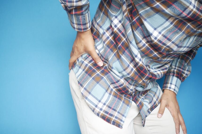
There is no correspondence between the anatomopathological condition and functional manifestations. Pain can start subtly in the iliac fossa and is exacerbated by thigh flexion. It does not cause prolonged functional impotence but visibly reduces sports performance. Sometimes, you can hear a "click" coming from the joint due to the hypo-sensitivity of the psoas and excessive tension on the corresponding tendons acting like a guitar string.
Rehabilitation treatment consists of myofascial massage of the iliopsoas muscle, recovery of extension and muscle strength, together with an evaluation of the piriformis muscle and a reflexogenic massage. Prior, it is necessary to rule out the possibility of joint blockage and misalignment of the sacrum.
You will be able to return to sports in a short time after a specific and personalized treatment. Iliopsoas injury causes significant functional limitation, leading to a state of concern and caution, especially because it is often a muscle injury that is not diagnosed when first aid is sought.
Conservative treatment is the only type of treatment suggested when this type of pathology occurs. Medical recovery includes: cessation of sports activities during the treatment period, usually requiring a minimum of 3 months, local analgesics, physiotherapy, TECAR WINBACK therapy, laser, myofascial massage, specific stretching, osteopathic techniques for repositioning the sacrum and pelvis. To decide when the patient can return to sports, variables such as the sports activity and the nature of the injury must be considered.
.jpg)
Road accidents are the main cause of pelvic fractures. These can be divided into three main groups:
You may face long periods of hospitalization with associated problems, neurological or cardiovascular. When you come to us, the physiotherapy plan will always be established after consultation with the surgeon who performed the operation, and the recovery will be gradual and progressive.
It should be noted that possible complications involving the nervous system may occur. These can be early and manifest immediately or later with more serious problems. Generally, a nervous system injury occurs in 1-5% of pelvic fractures cases. The percentage increases to 18% if both pelvic arches are affected, reaching up to a maximum of 33% when posterior acetabular fractures are accompanied by hip dislocation.
The sciatic nerve is the most frequently involved in these events. The recovery period usually lasts from 4 to 6 months, although it is subject to certain variables, especially advanced age.
.jpg)
The condition falls into the area of degenerative pathology, involving the wear processes of the joint heads, the so-called arthrosis. These degenerative processes lead to an attempt to heal the area and manage the problem. Unfortunately, these repairs are not entirely successful, and osteophytes (bone spurs) appear, which become an important factor in limiting joint mobility. Arthrosis can occur in both healthy and traumatized or malformed joints. Degeneration is more frequently encountered in women after menopause and in overweight individuals. Also, poor posture acquired at work can favor the pathology.
The symptoms are clear and include groin pain (which tends to worsen while walking or trigger after a period of inactivity), radiating towards the knee. This is why any patient complaining of knee pain will be examined at the hip joints level, especially when there is no history of knee trauma, weakness, loss, or imbalance.
The diagnosis is clinical and radiological, with X-rays used to discover bone structure changes, and CT and MRI to detect the presence of cartilage irregularities.
An appropriate rehabilitation plan aims to reduce pain, increase the range of motion, return the patient to daily active life, and most importantly, slow the disease's progression. The physiotherapy program is effective only when carried out early and correctly. It must be taken into account that joints are movement organs, and when one does not function properly, the body's natural response is to protect the damaged areas, especially those involved in movement.
Therefore, rehabilitation exercises will aim to restore the range of motion, increase muscle strength, and reduce imbalances. Wrong posture behaviors must be eliminated and good coordination maintained. These exercises can significantly help in pain control and improving the quality of life.
The patient may complain of mild pain in the sacroiliac joint, which can worsen when performing certain movements, therefore, limiting sports activities is necessary. Sometimes, the patient may also complain of a "click" or "clunk" noise coming from the hip.
Usually, there is evidence of an acute trauma or older microtrauma responsible for a painful contracture, simulating a joint problem at the hip or visceral pain, leading the patient to undergo a series of internal consultations. The pain can radiate to the groin area. In athletes, the iliopsoas muscle and tendon are usually responsible for many cases of groin pain – pubalgia. The iliopsoas or adductor muscles of the thigh are most often responsible for the pain and discomfort felt in this area.
The diagnosis is usually clinical and based on muscle tests to determine resistance and palpation. Sometimes, additional examinations are necessary to exclude a possible muscle injury (MRI scan) or involvement of the coxofemoral joint (X-ray).
The treatment is universally conservative and based on specific myofascial massage, in addition to relaxing muscle massage – on other muscle groups usually involved, posture and stretching exercises, selective toning of the psoas and synergistic muscles (muscle groups that contract together to perform the same body movement).
It is also very important to integrate manual therapies that can reposition any joint dysfunctions.
.jpg)
The patient may complain of a mild pain in the sacroiliac joint, which can worsen when performing certain movements; therefore, it is necessary to limit sports activities. Sometimes, the patient may complain of a "click" or "clunk" noise coming from the hip.
There is usually evidence of an acute trauma or older microtrauma responsible for a painful contracture, which simulates a hip joint problem or visceral pain, leading to a series of internal consultations. The pain may radiate to the groin area.
In athletes, the iliopsoas muscle and tendon are usually responsible for many cases of groin pain - pubalgia. The iliopsoas or thigh adductors are most often responsible for the pain and discomfort felt in this area.
The diagnosis is usually clinical and based on palpation and muscle tests to determine resistance. Sometimes, additional examinations are necessary to exclude possible muscle injury (MRI scan) or involvement of the coxofemoral joint (X-ray).
Treatment is universally conservative and consists of specific myofascial massage, relaxation, posturing, stretching exercises, selective toning of the psoas muscle, and synergistic muscles (groups of muscles that contract together to perform the same body movement).
It is also very important to integrate manual therapies, which can reposition any joint dysfunctions.
.jpeg)
When hip pain is severe and there are serious limitations in joint function, a complex X-ray can help the specialist suggest surgical intervention and replacement of the joint with a prosthesis.
This type of surgery is mainly recommended for people over 60 years old, considering the lifespan of the prosthesis and the decrease in physical activity in this age group.
Hip replacement allows for the recovery of good quality of life and pain relief, with a prosthesis lifespan of more than 10 years in 90% of cases.
The hip prosthesis can be of 4 types:
Rehabilitation after hip replacement surgery aims to recover joint movement, muscle strength, coordination, and walking ability. Your doctor will arrange for the start of rehabilitation in the hospital during the first days after surgery, with limb mobilization assisted by a therapist.
The period following hospital discharge is used to regain strength, mobility, and typical functionality of this area, with a remarkable commitment from both you and your physiotherapist.
You will be guided in both the physiotherapy room and the pool, where recovery is particularly rapid, followed by specific work to recover gestures that were no longer performed correctly.
See here how you can make an appointment and the location of our clinics.

CENTROKINETIC FLOREASCA
Bucharest, 1st District,
Mircea Eliade Street, no. 18,
Eliade Tower, Entrance A, Floor 3
| Navigation | Explore 3D |
CENTROKINETIC Barbu Vacarescu
Bucharest, 2nd District,
Barbu Vacarescu Street, no. 102,
102 The Address, ground floor.
| Navigation | Explore 3D |
CENTROKINETIC MILITARI
Bucuresti, Sector 6,
Bld. Iuliu Maniu, nr. 15G,
Cladirea PBT, etaj 3.
| Navigation |
CENTROKINETIC CLUJ-NAPOCA
Cluj-Napoca,
Str. Intre Lacuri, nr.1
Intrarea este in spatele cladirii.
| Navigation |
SOCRATES CLINIC TIMISOARA
Timisoara, Calea Martirilor 1989,
nr. 1.
Telefon: 0371 785 374.
| Navigation |
Phone: 0319693
E-mail: programari@centrokinetic.ro
Operating hours:
Monday – Friday: 07:00 – 21:00
Saturday: 09:00 – 14:00
Sunday: closed.
MAKE AN APPOINTMENT


