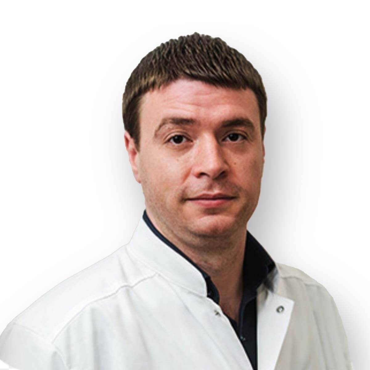
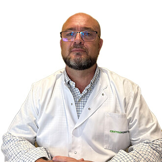
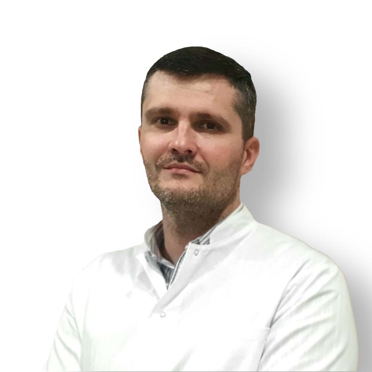
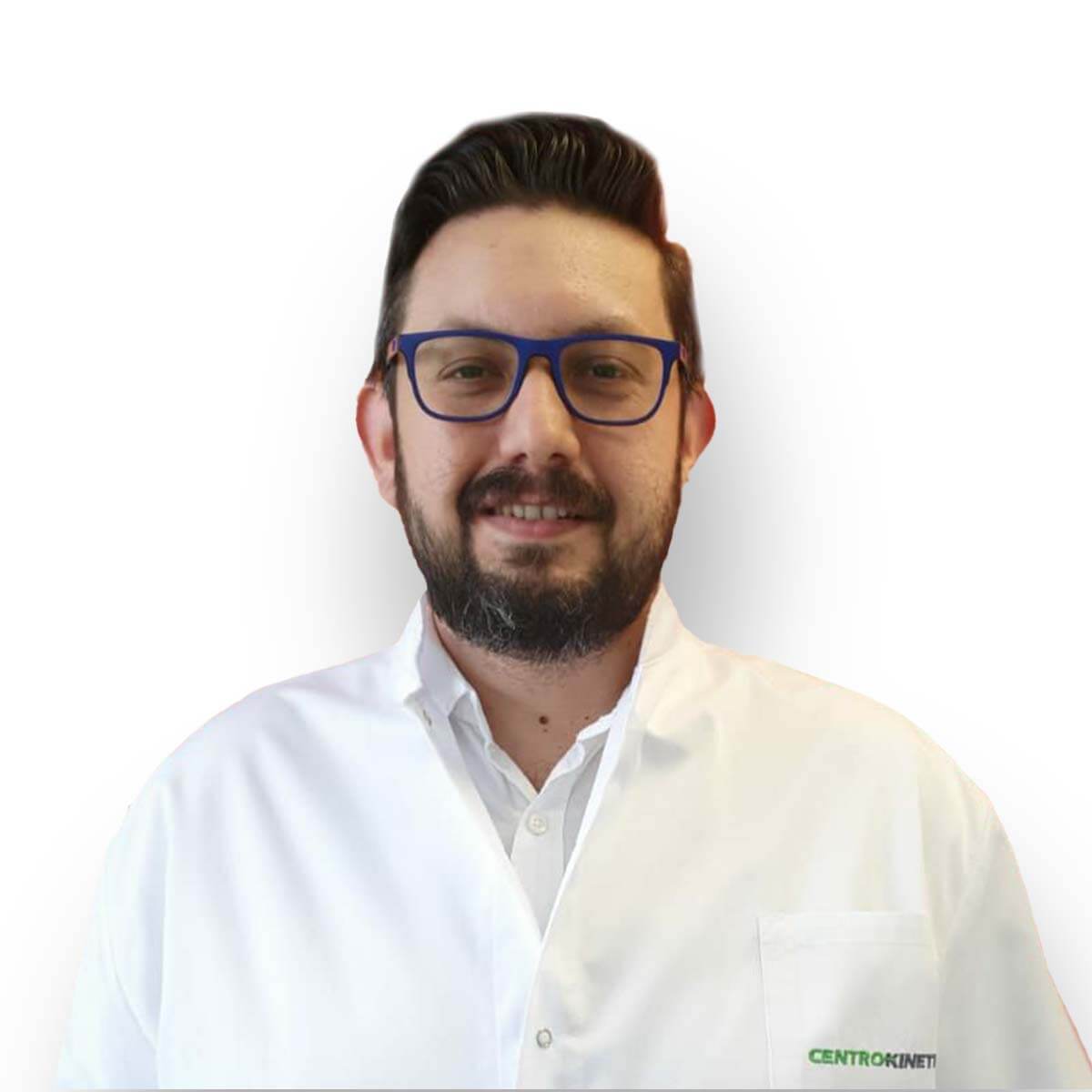
.jpg)
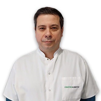
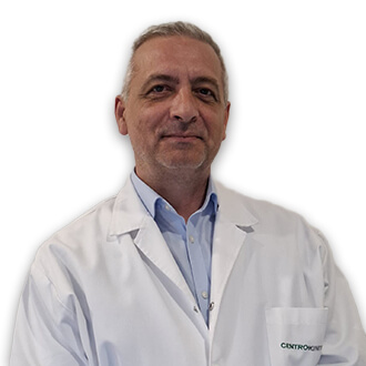
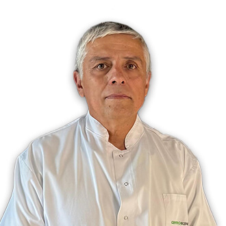
For all traumatic or chronic diseases of the musculoskeletal system, the Centrokinetic private clinic in Bucharest is prepared with an integrated Orthopedic Department, which offers all the necessary services to the patient, from diagnosis to complete recovery.
The Department of Orthopedic Surgery of Centrokinetic is dedicated to providing excellent patient care and exceptional education for young physicians in the fields of orthopedic surgery and musculoskeletal medicine.
Centrokinetic attaches great importance to the entire medical act: investigations necessary for correct diagnosis (ultrasound, MRI), surgery, and postoperative recovery.
Discover the open MRI imaging center in our clinic. Centrokinetic has a state-of-the-art MRI machine, dedicated to musculoskeletal conditions, in the upper and lower limbs. The MRI machine is open so that people suffering from claustrophobia can do this investigation. The examination duration is, on average, 20 minutes.
The shoulder joint offers such a large area of movement and much greater flexibility than other joints in the body, being consequently the joint most prone to dislocations and subluxations. Recurrent trauma, scapulohumeral dislocations, or congenital laxity of the soft parts can lead to a chronic condition called joint instability.
The Bankart procedure is a surgical technique that tries to regain the stability of the shoulder. It is indicated in recurrent shoulder dislocations, but sometimes this technique is used as a first intention, after an acute scapulohumeral dislocation. The technique involves arthroscopic reinsertion of the capsulo-ligamentous elements from the anterior part of the shoulder joint, with the help of anchors.
The bony components of the shoulder joint are the humeral head and the glenoid cavity of the scapula. The stability of the joint is provided by the joint capsule and the glenohumeral ligaments. The articular capsule is a cylindrical, fibrous, loose sleeve, located around the bony ends, ensuring the necessary freedom of movement and the removal of the articular surfaces by about 20 mm. It consists of deeper longitudinal and circular surface fibers. The capsule starts at the scapula and is inserted on the head of the humerus so that the two humeral tubercles remain outside the joint cavity. The lower part of the capsule is thin and loose, this area generating an increased incidence of anteroinferior shoulder dislocations. Above the capsule is the supraspinatus muscle, anteriorly the subscapularis muscle, posterior infraspinatus, and small round muscles. There is a rotating headband around it. The joint capsule is crossed by the long head of the brachial biceps muscle, which passes through the joint and is inserted on the scapula.
Joint ligaments: are fibrous bands considered thickenings of the joint capsule. Among them, we mention the glenohumeral ligaments (upper, middle, lower) located on the anterior face of the joint, the coracohumeral ligament, the coracoacromial ligament.
Shoulder dislocation is the most common dislocation and in 96% of cases occurs as a result of trauma. The anterior dislocation occurs through the mechanism of abduction, retroflexion, and forced external rotation of the arm, practically, by a fall on the hand with the arm in this position. The antero-internal variety is the most common, while the posterior dislocation, produced by internal hyper rotation, adduction, and anteflexion is the rarest.
Glenohumeral dislocation has an incidence of 2% in the population and 7% in athletes. It can be anterior-inferior (90%), posterior (4%), inferior or erect (1%). In 1% of cases, it is associated with a fracture of the humeral head or glenoid cavity. Shoulder dislocation always involves a rupture of the anterior capsule of the shoulder, more precisely a disinsertion at the level of the anterior edge of the glenoid cavity, both of the capsule and the glenohumeral ligaments.
The surgical technique can be performed both openly and arthroscopically, but in the last decade the minimally invasive technique has taken on a very large scale, presenting numerous advantages: 3 incisions of 1 cm, minimal complications, rapid recovery, hospitalization of 1-2 days, lower costs.
The intervention is performed with general anesthesia (AG IoT) or loco-regional anesthesia (interscalenic or supraclavicular block).
Subsequently, the patient is positioned either in the "beach chair position" or in lateral decubitus, the decision belonging to the orthopedist. An incision of about 1 cm is made in the posterior part of the shoulder joint at 1.5-2 cm lower and 1 cm medial from the postero-lateral angle of the acromion. It is the first portal and is very important because it inserts the arthroscope into the joint and into the subacromial space. Subsequently, these 2 spaces are inspected and the discovered lesions are counted. Then, the anterior portal is made: it is placed as standard between the brachial biceps, the subscapular ms, and the humeral head. This portal can be made from inside to outside or vice versa. Care must be taken with the musculocutaneous nerve, located 1 cm medially and 3 cm distal from the coracoid process. The external technique is performed with a needle, which establishes the ideal position of the portal. The last portal is the anteroinferior one: it can be performed from the outside inside or vice versa. It passes through the lower 1/3 of the subscapular ms, between the musculocutaneous and axillary nerves. It is used to repair previous shoulder instabilities. Subsequently, the anterior edge of the glenoid cavity must be prepared for reinsertion. That is why a special tool is used to produce bleeding, this bleeding being very important for healing.
.jpg)
Subsequently, wider threads or bands are passed through the joint capsule and glenohumeral ligaments for anchoring. The effective reinsertion of the capsulo-ligamentous elements to the bone is done with the help of anchors. Our medical team uses Arthrex anchors: PushLock or 4.5mm SwiftLock. Generally, you need 2, a maximum of 3 anchors.
At the end of the operation the upper limb is placed in a sling (immobilization). The intervention lasts, on average, 90 minutes. Subsequently, the patient is given antibiotics, painkillers, and anti-inflammatory drugs.
.jpg)
Post surgery
After the intervention, the patient remains hospitalized for 1-2 days. He will receive pain medication and antibiotics during his hospitalization. The operated limb is partially immobilized in a Dessault bandage for a few days.
After the operation, you will be discharged, with the related indications related to the recovery and the subsequent controls. Passive early mobilization of the shoulder joint is necessary for the patient to regain normal mobility. Our medical team guides the patient to physical therapy and physiotherapy under the guidance of one of our doctors.
The patient must understand that after the operation he has certain limitations in mobility, ie he is forbidden to make certain movements.
At home
Although recovery from arthroscopy is much faster than a classic operation, it will still take a few weeks for you to fully recover your shoulder joint. You should expect pain and discomfort for at least a week postoperatively. Ice will reduce pain and inflammation.
You must be careful not to sleep on the operated shoulder in the first weeks because the pain and discomfort can worsen. You can take a bath, but without wetting the bandage and incisions. The threads are suppressed at 14 days postoperatively. Physical therapy plays a very important role in the rehabilitation program, and the exercises must be supervized by a physical therapist until the end of the recovery period.
It is very important to follow the recovery program strictly and seriously for the surgery to be a success. Our medical team works on average with the patient after this intervention, 12-18 weeks until the complete recovery of the shoulder.

MAKE AN APPOINTMENT
CONTACT US
MAKE AN APPOINTMENT
FOR AN EXAMINATION
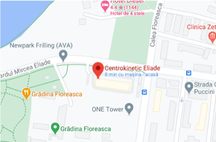
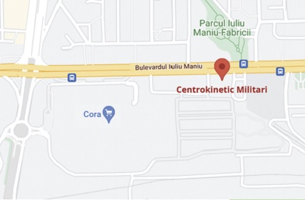
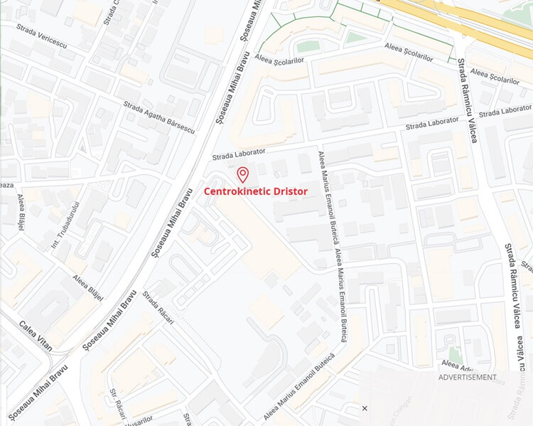
See here how you can make an appointment and the location of our clinics.
MAKE AN APPOINTMENT

