
.jpg)
Discover the open MRI imaging center in our clinic. Centrokinetic has a state-of-the-art MRI machine, dedicated to musculoskeletal conditions, in the upper and lower limbs. The MRI machine is open so that people suffering from claustrophobia can do this investigation. The examination duration is, on average, 20 minutes.
Centrokinetic attaches great importance to the entire medical act: investigations necessary for correct diagnosis (ultrasound, MRI), surgery, and postoperative recovery.
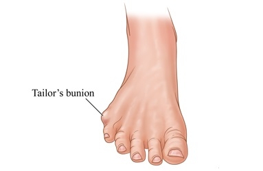
The Taylor's bunion is similar to the deviation of the first toe (hallux valgus), but develops on the outside of the foot, at the base of the fifth toe, at the metatarsophalangeal joint of the V ray. The name has its origins in the custom of tailors they stood cross-legged all day and this causes this deformity to appear on the outside of the foot.
CAUSE
The tailor's mount appears as a swelling at the base of the fifth toe, due to a high coefficient of friction inside shoes that are too narrow. The constant pressure produces a callus - a thickening of the tissues, which leads to the appearance of a painful mount on the outside of the foot. The affected tissues are inflamed and painful, and the use of inappropriate shoes accentuates the condition. If there is a bone deformity, then the situation can be even worse in terms of symptoms and treatment. The solution to reducing pain is to eliminate the pressure. The pressure can be reduced from the outside by changing the narrow shoes, or from the inside - surgically.
.jpg)
Symptoms
Symptoms include local inflammatory signs: pain, swelling, redness of the skin, sensitivity to palpation. Patients complain of difficulty in purchasing shoes that do not cause pain around the deformity. Local swelling causes a visible prominence, which some people consider unsightly.
Surgical technique
Under spinal anesthesia, an incision of about 2-3 cm is made at the base of the thumb, hemostasis and the swollen tissues are highlighted, respectively the joint capsule. If the prominence isn't noticeable, the surgical technique involves resection of the affected tissues, and the reconstruction of the joint capsule, the subcutaneous tissue, and the skin.
If the deviation is significant, the osteotomy of metatarsal V is performed and its fixation in the correction position, with a 2mm Herbert screw. The implant is designed to stay in place all the time. At any time, the patients can undergo MRIs.
.jpg)
Postoperative
After the operation, the patient remains hospitalized for 1 day. He will receive pain medication and antibiotics during hospitalization. The operated limb is not immobilized, and the patient is advised to make ankle exercises, from the first postoperative day. Free walking is allowed immediately, without the need for crutches, given that the chosen intervention was joint debridement.
.jpg)
In the case of arthrodesis, patients will wear a compressive bandage on the foot for 5 days and will use a special shoe when walking, which correctly distributes the body weight. Patients can return to daily activities quickly, up to 3-4 weeks.
At home
Although recovery after this operation is much faster than a classic intervention, it will still take a few weeks for you to fully recover the operated joint. You should expect pain and discomfort for at least a week postoperatively. You can use a special ice pack, which will reduce the pain and inflammation. You must be careful not to lean on the operated area in the first weeks because the pain and discomfort can worsen. You can take a bath, but without wetting the bandage and incisions. The threads are suppressed at 14 days postoperatively.
At 6 weeks postoperatively, an X-ray is necessary to see how the affected joint heals. Driving is allowed after 6 weeks, and hard physical work after 12 weeks.
Physical therapy plays a very important role in the rehabilitation program, and the exercises must be followed by a physical therapist until the end of the recovery period.
It is very important to follow the recovery program strictly and seriously for the surgery to be a success. Our medical team works on average with the patient after this intervention, 18-24 weeks until complete recovery of the operated area.
Following any surgery, medical recovery plays an essential role in the social, professional, and family reintegration of the patient. Because we pursue the optimal outcome for each patient entering the clinic, recovery medicine from Centrokinetic is based on a team of experienced physicians and physical therapists and standardized medical protocols.
See here how you can make an appointment and the location of our clinics.

CENTROKINETIC FLOREASCA
Bucharest, 1st District,
Mircea Eliade Street, no. 18,
Eliade Tower, Entrance A, Floor 3
| Navigation | Explore 3D |
CENTROKINETIC Barbu Vacarescu
Bucharest, 2nd District,
Barbu Vacarescu Street, no. 102,
102 The Address, ground floor.
| Navigation | Explore 3D |
CENTROKINETIC MILITARI
Bucuresti, Sector 6,
Bld. Iuliu Maniu, nr. 15G,
Cladirea PBT, etaj 3.
| Navigation |
CENTROKINETIC CLUJ-NAPOCA
Cluj-Napoca,
Str. Intre Lacuri, nr.1
Intrarea este in spatele cladirii.
| Navigation |
SOCRATES CLINIC TIMISOARA
Timisoara, Calea Martirilor 1989,
nr. 1.
Telefon: 0371 785 374.
| Navigation |
Phone: 0319693
E-mail: programari@centrokinetic.ro
Operating hours:
Monday – Friday: 07:00 – 21:00
Saturday: 09:00 – 14:00
Sunday: closed.
MAKE AN APPOINTMENT


