
FOOT AND ANKLE
The Foot and Ankle
The foot has a strong and complex structure: it is composed of 26 bones, connected by 33 joints and strengthened by more than 100 ligaments. The body weight is transmitted to the foot through the ankle or the tibio-tarsal joint. The tibia and fibula with the two malleoli surpass and surround the upper part of the talus, which allows the transfer of pressure to the other bones of the foot.
Functions of the Foot
- Represents the base of the body and provides the necessary support for an upright position using minimal effort
- Ensures the necessary torsion mechanism for the tibia and fibula during the loading phase of walking
- Provides elasticity to adapt to different surfaces in walking or running
- Absorbs intermittent shocks
With the main role of supporting the body in an upright position and propelling it during walking, the ankle and foot are prone to various degenerative conditions and injuries.
Contents
Common Conditions
- ankle sprain
- metatarsal fracture
- malleolar fractures
- cartilage injuries
- Achilles tendon rupture
- Lisfranc joint fracture/injury
Chronic Conditions
- Achilles tendon disorders
- plantar fasciitis and heel spurs
- Morton's neuroma
Surgical Interventions
- malleolar fractures
- cartilage injuries
- bunions – hallux valgus
Common Conditions
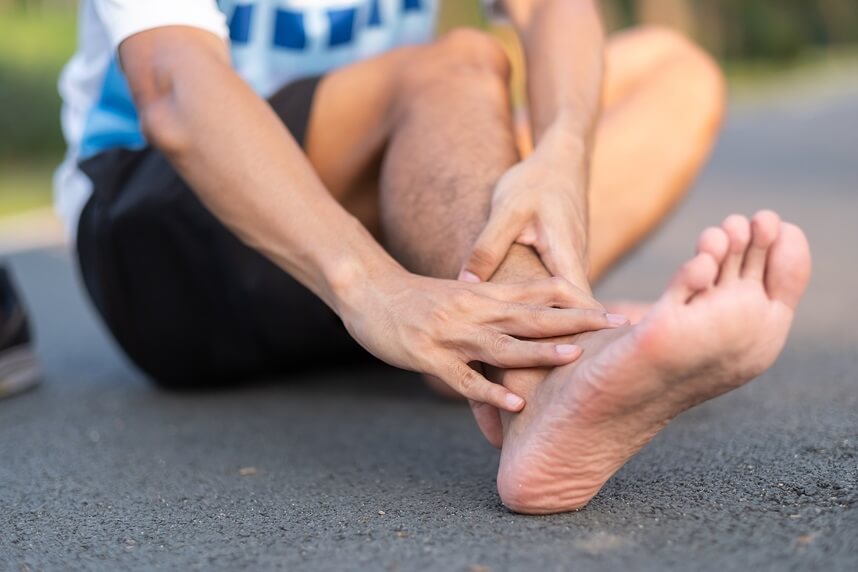
- ankle sprain
.jpg)
Most people will suffer an ankle sprain during their lifetime, this being the most common injury with a higher prevalence among athletes.
The most common cause is the internal rotation of the foot (inversions). Sprains can also result from an eversion (an external rotation of the foot) and sometimes the two injuries can coincide. The ligament most prone to an inversion is the anterior talofibular, followed by the calcaneofibular and then the posterior talofibular. Meanwhile, eversions are more often the result of an injury to the deltoid ligament.
Swelling usually appears immediately, and the pain can be very intense. Movements are severely limited due to the swelling, while ankle stability can also be compromised.
If the area around the ankle is remarkably swollen, an X-ray is taken to rule out the suspicion of a fracture. An ultrasound performed a few days after the injury can help highlight deformations and ligament injuries. In rare cases, the examination may be completed with an MRI or CT scan.
Rehabilitation following acute traumatic injuries is essential for pain relief, restoring joint stability for dynamic function. It is also important for the patient to continue a maintenance program after rehabilitation is complete to avoid the recurrence of the problem.
- metatarsal fracture
.png)
Fractures of the fourth and fifth metatarsal are the most common and usually do not require surgery. Patients usually present with pain following a sprain or after a fall or jump. Motorcycle or car accidents are also responsible for these injuries.
The normal course of treatment involves immobilizing the foot with a cast for approximately 30 days, during which muscle atrophy in the foot will be evident. Again, X-rays are extremely important for diagnosis. Once the therapist has verified the consolidation of the fracture and the correct juxtaposition of the bone ends, rehabilitation can begin.
The rehabilitation program will start with very light loading and will progress steadily until crutches are no longer needed. Before field rehabilitation can begin, the patient must have a wide range of motion, fluidity, proprioception, and good muscle strength in the lower limb.
If osteosynthesis techniques have been used, the rehabilitation program does not change significantly, although resistance exercises can start earlier, reducing the overall duration of the process.
- malleolar fractures
.jpg)
These types of fractures can be caused by a wide range of incidents, such as car accidents, sports injuries, and even accidents at home or work. These injuries will normally require immobilization of the joint with a cast or brace for 30-40 days.
After this period, the ankle will be extremely stiff in all planes (flexion-extension and inversion-eversion), and muscle hypotrophy will be evident.
If you have suffered this type of injury and come to us for rehabilitation, it is very important to bring X-rays of the area with you, especially those taken after the cast has been removed. These X-ray films inform your doctor whether your malleolar bones are aligned – if they are not, successful rehabilitation becomes impossible.
Rehabilitation of these types of injuries can require a long period of time. Physical and pharmacological strategies will be used to reduce pain and swelling, and, in addition, manual lymphatic drainage is often necessary. Later, during therapy, proprioceptive exercises will be introduced in parallel with progressive loading to help strengthen the muscles around the ankle.
Aquatic rehabilitation (hydrotherapy) is very effective when dealing with such injuries, as it allows the patient to regain walking with minimal load.
These injuries can particularly disrupt the patient's life due to immobilization of the foot. We try to help our patients achieve the basic movements necessary to return to normal life (helping them to drive, walk without crutches, and eventually return to sports or their usual activities).
.jpg)
These injuries most often occur as a result of a deformity during supination, usually affecting the medial compartment, resulting in persistent pain and functional limitation.
Patients (usually professional athletes) report numerous ankle sprains, persistent pain, limited movements, and swelling.
Varus-valgus and drawer tests may have positive results in these cases. X-rays are not precise enough to be used. MRI provides better visualization of both osteochondral fractures and any dislocation of bone fragments.
Treatments for these conditions vary depending on their severity. As long as it is not a grade 4 injury (one that includes dislocated fragments, which can only be remedied surgically), treatment based on conservative rehabilitation can begin.
.jpg)
The Achilles tendon is the largest and most robust tendon in the human body.
Repetitive stress in athletes or simple aging in inactive individuals can lead to variations in the structure of this tendon. This can lead to partial or even complete ruptures of the tendon. These conditions often result from unrecognized or misunderstood chronic tendinitis. It mainly affects runners, soccer players, and tennis players. It is believed to result from a sudden contraction.
Athletes often state that they felt as if they had been kicked by an opponent. The rupture generates immediate functional impairment, preventing any kind of movement.
The diagnosis is mainly based on the clinical presentation: sometimes there is an obvious gap at the rupture site. The suspected diagnosis is usually confirmed by an ultrasound that clearly shows the interruption of the tendon fibers and allows us to distinguish between partial and total ruptures.
In these cases, surgical intervention is always necessary.
Recovery is long-term. After a certain period of post-surgical immobilization, tendon loading is done progressively, with full walking recovery in a few months.
.jpg)
This injury is rare and difficult to diagnose and involves the tarsometatarsal joint (also known as the Lisfranc joint). It can occur through both direct and indirect mechanisms.
A classic example of direct trauma is a direct lateral or medial blow to the midfoot, which is usually unnoticed or underestimated by the patient. Indirect traumas include falls or getting the bare foot stuck in awkward places.
Symptoms are characterized by intense pain localized in the midfoot, which then becomes exaggerated along the Lisfranc joint line during compression.
About 20% of Lisfranc dislocation X-rays show negative results, but CT is very effective in highlighting signs indicating this type of injury.
Usually, patients suffering from this type of injury, but who do not present instability, are given a brace for 4-6 weeks, during which remission signs begin to appear. Physiotherapy is used to reduce pain and inflammation. A rehabilitation program is developed to help strengthen the ankle muscles and improve the cavity position along the plantar arch.
When dealing with unstable injuries, surgical treatment is most often necessary, consisting of percutaneous pinning using wires or screws, followed by immobilization with a brace or cast. Subsequent rehabilitation techniques, after the cast is removed, follow the same principles as in stable injuries.
Chronic Conditions
- Achilles tendon disorders
.jpg)
This category includes several inflammatory and degenerative conditions, including tendinitis, tendinosis, and insertional tendinitis (enthesitis). They can be the result of acute injury, triggered by functional overload or microtraumas caused by improper footwear, intense training, or exercises performed at low temperatures.
Initially, symptoms tend to worsen during rest, then subside after movement (the first steps in the morning can be particularly uncomfortable). After a while, the pain may no longer subside after movement, severely impeding the patient's walking. Over time, stress in the distal portion of the tendon can lead to inflammation of the pre-Achilles bursa, further complicating the clinical situation.
The diagnosis is based on the location of the pain (usually around the tendon insertion on the calcaneus), as well as inflammation and redness of the skin. These indicators are usually complemented by an ultrasound, used to accurately determine the location and extent of the injury.
Tendinopathies require delicate treatment, and the chances of success vary depending on the severity of the injury and the time elapsed since the onset of the first symptoms. However, forming an effective rehabilitation plan is important, as even the later, less severe stages of this condition should not be ignored.
- plantar fasciitis and heel spurs
.jpg)
Plantar fasciitis is a disorder concerning the fibrous connective tissue structures that originate from the calcaneal tuberosity and insert into the metatarsal heads.
During the support phase of walking and running movements, the plantar fascia is significantly stretched, causing a high level of stress around its insertion on the medial calcaneal tubercle. Over time, calcification can occur along the length of this band, resulting in the formation of spurs. The presence of these spurs is not necessarily linked to the presence of pain, as spurs are often discovered incidentally during X-rays for other injuries that have not caused the patient any pain, while some patients present very painful plantar fasciitis in the absence of a spur.
This condition is common among athletes involved in running, dancing, tennis, and basketball, especially if they have increased the load too quickly during training.
It can also affect older people who have started wearing flat shoes, those who are overweight, people who have to wear inappropriate shoes at work, and those with foot irregularities (flat feet, hard soles, and those with a tendency to overpronation). Generally speaking, this condition tends to be chronic, as it is often overlooked or neglected by sufferers for several months, during which it worsens.
Symptoms usually consist of severe pain during the first steps of the day, which gradually improves after some movement before worsening again later. Swelling may also be present around the affected area. It is not uncommon for patients to experience deficits in strength and extensibility.
X-rays, ultrasound, and possibly electromyography are useful for establishing a diagnosis. Electromyography is particularly useful if there is any numbness or paralysis due to compression of the associated nerve.
In the short term, the patient should stop any sporting activity (except swimming and cycling) and try to reduce the effect of predisposing factors (avoiding wearing inappropriate shoes or losing weight, for example). The use of insoles can help correct any arch abnormalities. Shockwave therapy can be very effective in reducing inflammation in these conditions.
- Morton's neuroma
.jpg)
Morton's neuroma is often attributed to swelling in the branches of the plantar nerve located between the second and third, or third and fourth metatarsals. Compression of these nerves between the metatarsal heads can be caused by microtraumas resulting from the use of inappropriate footwear (especially narrow shoes).
Patients usually present with a sudden onset of pain, often compared to an electric shock. Numbness is also frequently present in the two affected toes.
The diagnosis is essentially clinical but can be confirmed by an ultrasound or MRI scan. Initial treatment is conservative, but in severe cases where surgical intervention is necessary, the neuroma is removed.
Surgical Interventions
- malleolar fractures
.jpg)
These are the most common fractures of the lower limb: they involve the internal and external malleoli, sometimes combined with injuries to the ankle ligaments. A fracture involving at least two malleoli and the posterior part of the tibia is defined as a tri-malleolar fracture.
You have likely suffered trauma from a car accident, an injury caused during a sporting activity, or an accidental fall. Depending on the different types of fractures, there are various surgical treatments you may undergo.
Numerous synthetic bone substitutes are used just as well as external fixators. You will be able to start rehabilitation after a certain period of immobilization with a plaster or fixator. It is important to know that the proposed rehabilitation for this type of pathology is long-term and challenging, requiring an average of four months to achieve a modest recovery of functionality and approximately eight months to return to sports activity. In general, synthetic bone substitutes are removed one year after surgery. Rehabilitation after their removal will take almost a month.
- cartilage injuries
.png)
Cartilaginous injuries are often observed as a result of joint wear due to repetitive movements, although they are just as common as a result of a sports injury. Treatment in this case can be conservative or surgical in more severe cases. Either way, we can effectively treat this type of injury using our own conservative rehabilitation process or by developing a specialized post-surgical rehabilitation program.
- bunions – hallux valgus
.jpg)
This is an "acquired deformity" of the metatarsophalangeal joint of the big toe. A month after the corrective surgery, you will usually be advised to wear a wedge-shaped shoe that allows rocking from the heel (Talus shoe). Then, you will be able to start the rehabilitation process.
Pain will be present during the early stages after surgery, especially when some pressure acts on the scar (especially at night) and swelling will affect the front part of the foot.
Recovery begins with physical therapies to reduce pain and swelling, lymphatic drainage, and foot massage; then mobilizing the ankle and big toe joint and gradually strengthening the foot, calf, and thigh muscles, exercises to prepare for walking, and gradually abandoning crutches.
BUCHAREST TEAM


CLUJ NAPOCA TEAM


BRASOV TEAM


MAKE AN APPOINTMENT
FOR AN EXAMINATION
See here how you can make an appointment and the location of our clinics.
MAKE AN APPOINTMENT
Asiguratori privati

Parteneriate
CENTROKINETIC FLOREASCA
Bucharest, 1st District,
Mircea Eliade Street, no. 18,
Eliade Tower, Entrance A, Floor 3

| Navigation | Explore 3D |
CENTROKINETIC Barbu Vacarescu
Bucharest, 2nd District,
Barbu Vacarescu Street, no. 102,
102 The Address, ground floor.

| Navigation | Explore 3D |
CENTROKINETIC MILITARI
Bucuresti, Sector 6,
Bld. Iuliu Maniu, nr. 15G,
Cladirea PBT, etaj 3.

| Navigation |


CENTROKINETIC CLUJ-NAPOCA
Cluj-Napoca,
Str. Intre Lacuri, nr.1
Intrarea este in spatele cladirii.

| Navigation |


SOCRATES CLINIC TIMISOARA
Timisoara, Calea Martirilor 1989,
nr. 1.
Telefon: 0371 785 374.

| Navigation |
CONTACT
Phone: 0319693
E-mail: programari@centrokinetic.ro
Operating hours:
Monday – Friday: 07:00 – 21:00
Saturday: 09:00 – 14:00
Sunday: closed.
MAKE AN APPOINTMENT

